npj Parkinson's Disease ( IF 6.7 ) Pub Date : 2025-02-11 , DOI: 10.1038/s41531-025-00872-w
Matteo Santoro, Rachel K. Lam, Sarah E. Blumenfeld, Weiqi Tan, Peter Ciari, Emily K. Chu, Nay L. Saw, Daniel Ryskamp Rijsketic, Jennifer S. Lin, Boris D. Heifets, Mehrdad Shamloo
|
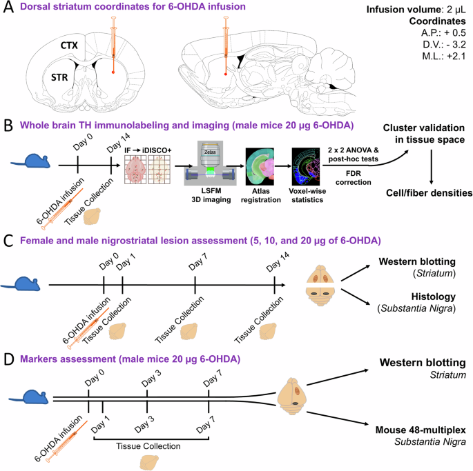
Efforts to develop disease-modifying treatments for Parkinson’s disease (PD) have been hindered by the lack of animal models replicating all hallmarks of PD and the insufficient attention to extra-nigrostriatal regions pathologically critical for the prodromal appearance of non-motor symptoms. Among PD models, 6-hydroxydopamine (6-OHDA) infusion in mice has gained prominence since 2012, primarily focusing on the nigrostriatal region. This study characterized tyrosine hydroxylase-positive neuron and fiber loss across the brain following a unilateral 6-OHDA (20 µg) infusion into the dorsal striatum. Our analysis integrates immunolabeling, brain clearing (iDISCO+), light sheet microscopy, and computational methods, including fMRI and machine learning tools. We also examined sex differences, disease progression, neuroinflammatory responses, and pro-apoptotic signaling in nigrostriatal regions of C57BL/6 mice exposed to varying 6-OHDA dosages (5, 10, or 20 µg) followed by 1, 7, and 14 days of recovery. This comprehensive, spatiotemporal analysis of 6-OHDA-induced pathology was used to map the time course of neuronal degeneration and the onset of neuroinflammation.
中文翻译:
儿茶酚胺能去神经支配、神经变性和炎症在 6-OHDA 处理的帕金森病小鼠中的图谱
由于缺乏复制 PD 所有特征的动物模型以及对非运动症状的前驱症状至关重要的黑质纹状体外区域的关注不足,开发帕金森病 (PD) 的疾病修饰疗法的努力受到了阻碍。在 PD 模型中,自 2012 年以来,小鼠 6-羟基多巴胺 (6-OHDA) 输注越来越受到重视,主要集中在黑质纹状体区域。本研究表征了单侧 6-OHDA (20 μg) 输注到背侧纹状体后酪氨酸羟化酶阳性神经元和整个大脑的纤维丢失。我们的分析集成了免疫标记、大脑清理 (iDISCO+)、光片显微镜和计算方法,包括 fMRI 和机器学习工具。我们还检查了暴露于不同 6-OHDA 剂量 (5、10 或 20 μg) 的 C57BL/6 小鼠黑质纹状体区域的性别差异、疾病进展、神经炎症反应和促凋亡信号,然后恢复 1、7 和 14 天。这种对 6-OHDA 诱导的病理学的全面时空分析用于绘制神经元变性的时间进程和神经炎症的发作。


 京公网安备 11010802027423号