当前位置:
X-MOL 学术
›
Adv. Mater.
›
论文详情
Our official English website, www.x-mol.net, welcomes your
feedback! (Note: you will need to create a separate account there.)
Molecular Imaging of Ovarian Follicles and Tumors With Near‐Infrared II Bioconjugates
Advanced Materials ( IF 27.4 ) Pub Date : 2024-12-19 , DOI: 10.1002/adma.202414129 Yicong Wang, Wenhan Lu, Zi‐Han Chen, Yan Xiao, Yu Wang, Wenhao Gao, Zhiming Wang, Ruihu Song, Zhao Fang, Wei Hu, Xiaoyu Tong, Kuinyu Lee, Zhenle Pei, Minzhen Xu, Fan Zhang, Hao Chen, Yi Feng
Advanced Materials ( IF 27.4 ) Pub Date : 2024-12-19 , DOI: 10.1002/adma.202414129 Yicong Wang, Wenhan Lu, Zi‐Han Chen, Yan Xiao, Yu Wang, Wenhao Gao, Zhiming Wang, Ruihu Song, Zhao Fang, Wei Hu, Xiaoyu Tong, Kuinyu Lee, Zhenle Pei, Minzhen Xu, Fan Zhang, Hao Chen, Yi Feng

|
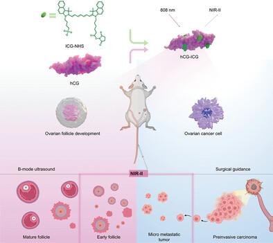
Follicular tracking is typically conducted using ultrasound technology, but its effectiveness is constrained by limited resolution. High‐resolution imaging of deep tissues can be accomplished using luminescence imaging in the near‐infrared II window (NIR‐II, 1000–1700 nm); however, the contrast agents that are used lack specificity. Here, it is reported that the FDA‐approved indocyanine green (ICG)‐conjugated recombinant human chorionic gonadotropin (hCG) protein can target early follicles with long‐term effectiveness. A novel high‐resolution NIR‐II imaging approach is developed for monitoring follicular development as well as ovulation using multi‐color imaging of ovarian vessels with a combination of non‐overlapping downconversion nanoparticles (DCNPs). The results showed that the ability to monitor early follicles of around 50 µm in diameter exceeded the spatial and temporal resolution of ultrasound or MRI without the reproductive damage associated with computed tomography radiation, and this enabled the clinical identification of the follicular reserve in patients with infertility diseases such as polycystic ovary syndrome (PCOS). In addition, NIR‐II imaging clearly targeted ovarian tumors and showed micro‐metastatic lesions, thus providing a new tool for monitoring tumors in vivo and guiding surgical resection.
中文翻译:

使用近红外 II 生物偶联物对卵巢卵泡和肿瘤进行分子成像
卵泡跟踪通常使用超声技术进行,但其有效性受到分辨率有限的限制。使用近红外 II 窗口 (NIR-II, 1000–1700 nm) 中的发光成像可以完成深部组织的高分辨率成像;然而,使用的造影剂缺乏特异性。据报道,FDA 批准的吲哚菁绿 (ICG) 偶联的重组人绒毛膜促性腺激素 (hCG) 蛋白可以长期有效地靶向早期卵泡。开发了一种新的高分辨率 NIR-II 成像方法,用于使用卵巢血管的多色成像和非重叠的下转换纳米颗粒 (DCNP) 的组合来监测卵泡发育和排卵。结果表明,监测直径约为 50 μm 的早期卵泡的能力超过了超声或 MRI 的空间和时间分辨率,而没有与计算机断层扫描辐射相关的生殖损伤,这使得能够临床识别不孕症患者的卵泡储备,例如多囊卵巢综合征 (PCOS)。此外,NIR-II 成像明确靶向卵巢肿瘤并显示微转移病灶,从而为体内监测肿瘤和指导手术切除提供了新工具。
更新日期:2024-12-19
中文翻译:

使用近红外 II 生物偶联物对卵巢卵泡和肿瘤进行分子成像
卵泡跟踪通常使用超声技术进行,但其有效性受到分辨率有限的限制。使用近红外 II 窗口 (NIR-II, 1000–1700 nm) 中的发光成像可以完成深部组织的高分辨率成像;然而,使用的造影剂缺乏特异性。据报道,FDA 批准的吲哚菁绿 (ICG) 偶联的重组人绒毛膜促性腺激素 (hCG) 蛋白可以长期有效地靶向早期卵泡。开发了一种新的高分辨率 NIR-II 成像方法,用于使用卵巢血管的多色成像和非重叠的下转换纳米颗粒 (DCNP) 的组合来监测卵泡发育和排卵。结果表明,监测直径约为 50 μm 的早期卵泡的能力超过了超声或 MRI 的空间和时间分辨率,而没有与计算机断层扫描辐射相关的生殖损伤,这使得能够临床识别不孕症患者的卵泡储备,例如多囊卵巢综合征 (PCOS)。此外,NIR-II 成像明确靶向卵巢肿瘤并显示微转移病灶,从而为体内监测肿瘤和指导手术切除提供了新工具。


 京公网安备 11010802027423号