当前位置:
X-MOL 学术
›
Anal. Chem.
›
论文详情
Our official English website, www.x-mol.net, welcomes your
feedback! (Note: you will need to create a separate account there.)
Cellular and Intravital Nucleus Imaging by a D-π-A Type of Red-Emitting Two-Photon Fluorescent Probe
Analytical Chemistry ( IF 6.7 ) Pub Date : 2024-12-17 , DOI: 10.1021/acs.analchem.4c04103 De-Chen Duan, Gaowei Pan, Junru Liu, Hao Chen, Tao Xie, Ying Long, Fang Dai, Shengxiang Zhang, Bo Zhou
Analytical Chemistry ( IF 6.7 ) Pub Date : 2024-12-17 , DOI: 10.1021/acs.analchem.4c04103 De-Chen Duan, Gaowei Pan, Junru Liu, Hao Chen, Tao Xie, Ying Long, Fang Dai, Shengxiang Zhang, Bo Zhou

|
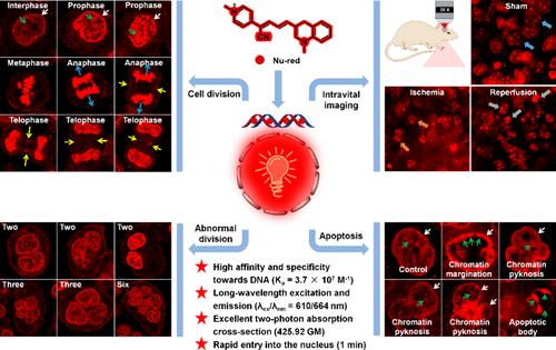
The advancement in fluorescent probe technology for visualizing nuclear morphology and nucleic acid distribution in live cells and in vivo has attracted considerable interest within the biomedical research community, as it offers invaluable insights into cellular dynamics across various physiological and pathological contexts. In this study, we present a novel two-photon nucleus-imaging fluorescent probe called Nu-red, which is a typical donor(D)-π-acceptor(A) rotor composed of the donor (dihydroquinoline) and acceptor (pyridiniumylpentadienitrile) parts linked by a single bond. This probe offers several advantages, including long-wavelength excitation and emission (λex/λem = 610/664 nm), favorable quantum yields (1.35–22.15%), excellent two-photon absorption cross-section (425.92 GM), high selectivity and sensitivity, high DNA-binding affinity (Ka = 3.7 × 10⁷ M–1, comparable to that of the commercial nucleus stain Hoechst 33342), rapid entry into the nucleus (1 min), low cytotoxicity, membrane-permeability, good water solubility, applicability to various cell lines, and compatibility with other commercial probes. Leveraging these aforementioned advantages, Nu-red was successfully employed to visualize cell division in living cells, distinguish abnormal division cells from normal ones, and track morphological changes in the nucleus during cell apoptosis. More notably, Nu-red was utilized to visualize nuclear shrinkage and pyknosis in the brain of a living mouse model of ischemic stroke.
中文翻译:

通过 D-π-A 型红光双光子荧光探针进行细胞核和活体细胞核成像
用于可视化活细胞和体内细胞核形态和核酸分布的荧光探针技术的进步引起了生物医学研究界的极大兴趣,因为它为各种生理和病理背景下的细胞动力学提供了宝贵的见解。在这项研究中,我们提出了一种名为 Nu-red 的新型双光子核成像荧光探针,它是一个典型的供体 (D)-π-受体 (A) 转子,由供体(二氢喹啉)和受体(吡啶基五二烯腈)部分组成,由单键连接。该探针具有多种优势,包括长波长激发和发射 (λex/λem = 610/664 nm)、良好的量子产率 (1.35–22.15%)、出色的双光子吸收截面 (425.92 GM)、高选择性和灵敏度、高 DNA 结合亲和力 (Ka = 3.7 × 10⁷ M–1,与商业细胞核染色剂 Hoechst 33342 相当)、快速进入细胞核(1 分钟)、低细胞毒性、膜通透性、良好的水溶性、适用于各种细胞系以及与其他商业探针的兼容性。利用上述优势,Nu-red 成功地用于可视化活细胞中的细胞分裂,区分异常分裂细胞和正常细胞,并跟踪细胞凋亡过程中细胞核的形态变化。更值得注意的是,Nu-red 被用于可视化缺血性中风活体小鼠模型大脑中的核收缩和 pyknosis。
更新日期:2024-12-17
中文翻译:

通过 D-π-A 型红光双光子荧光探针进行细胞核和活体细胞核成像
用于可视化活细胞和体内细胞核形态和核酸分布的荧光探针技术的进步引起了生物医学研究界的极大兴趣,因为它为各种生理和病理背景下的细胞动力学提供了宝贵的见解。在这项研究中,我们提出了一种名为 Nu-red 的新型双光子核成像荧光探针,它是一个典型的供体 (D)-π-受体 (A) 转子,由供体(二氢喹啉)和受体(吡啶基五二烯腈)部分组成,由单键连接。该探针具有多种优势,包括长波长激发和发射 (λex/λem = 610/664 nm)、良好的量子产率 (1.35–22.15%)、出色的双光子吸收截面 (425.92 GM)、高选择性和灵敏度、高 DNA 结合亲和力 (Ka = 3.7 × 10⁷ M–1,与商业细胞核染色剂 Hoechst 33342 相当)、快速进入细胞核(1 分钟)、低细胞毒性、膜通透性、良好的水溶性、适用于各种细胞系以及与其他商业探针的兼容性。利用上述优势,Nu-red 成功地用于可视化活细胞中的细胞分裂,区分异常分裂细胞和正常细胞,并跟踪细胞凋亡过程中细胞核的形态变化。更值得注意的是,Nu-red 被用于可视化缺血性中风活体小鼠模型大脑中的核收缩和 pyknosis。


 京公网安备 11010802027423号