当前位置:
X-MOL 学术
›
Anal. Chem.
›
论文详情
Our official English website, www.x-mol.net, welcomes your
feedback! (Note: you will need to create a separate account there.)
Gold Nanoparticle Detection with Two-Photon Excitation Fluorescence Lifetime Imaging of NAD(P)H in Cancer Cells: An Analytical Approach to Separate Nanoparticle and NAD(P)H Signals
Analytical Chemistry ( IF 6.7 ) Pub Date : 2024-12-12 , DOI: 10.1021/acs.analchem.4c04214 Mahshid Ghasemi, Amy Holmes, Tyron Turnbull, Ivan Kempson
Analytical Chemistry ( IF 6.7 ) Pub Date : 2024-12-12 , DOI: 10.1021/acs.analchem.4c04214 Mahshid Ghasemi, Amy Holmes, Tyron Turnbull, Ivan Kempson

|
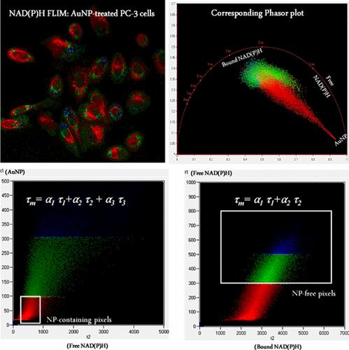
Gold nanoparticles (AuNPs) have shown promise for applications in the diagnosis and treatment of different diseases, including cancer. Understanding the effect of AuNPs on metabolic reprogramming in cancer cells at the single cell level is of high importance for improving the efficacy and safety. Fluorescence lifetime imaging microscopy (FLIM) of nicotinamide adenine dinucleotide (phosphate) hydrogen (NAD(P)H) as a main metabolic cofactor and an indicator of metabolic reprogramming in cancer cells enables real-time monitoring of cancer cell metabolism in response to different treatments, including AuNPs. However, NPs such as AuNPs can be a potential source of signals themselves, which provides opportunities to measure the NP internalization, but it is also important to minimize confounding effects on metabolic measurements. In this study, we detected inherent photoluminescence (PL) from the AuNPs in treated prostate cancer cells (PC-3 cell line) as well as in solution at the NAD(P)H emission wavelength. We developed an analysis approach to minimize the confounding effect of the AuNPs’ PL on metabolic measurements. On the other hand, we assessed the reliability of the intracellular AuNPs’ PL as an estimator of AuNP uptake. To assess if intracellular AuNPs’ PL may be dependent on the exposed cell type, we performed NAD(P)H FLIM imaging of AuNP-exposed SKBR-3 breast cancer cells, where we observed a similar AuNP PL but at a much lower level compared to PC-3 cells. We proposed that this difference can be attributed to the different levels of AuNP uptake or varying intracellular microenvironments.
中文翻译:

使用双光子激发荧光寿命成像对癌细胞中的 NAD(P)H 进行金纳米颗粒检测:一种分离纳米颗粒和 NAD(P)H 信号的分析方法
金纳米颗粒 (AuNPs) 已显示出在诊断和治疗包括癌症在内的不同疾病方面的应用前景。在单细胞水平上了解 AuNPs 对癌细胞代谢重编程的影响对于提高疗效和安全性非常重要。烟酰胺腺嘌呤二核苷酸(磷酸盐)氢 (NAD(P)H) 作为主要代谢辅助因子和癌细胞代谢重编程的指标的荧光寿命成像显微镜 (FLIM) 能够实时监测癌细胞代谢对不同治疗(包括 AuNPs)的反应。然而,NP (如 AuNPs) 本身可以是潜在的信号源,这为测量 NP 内化提供了机会,但尽量减少对代谢测量的混杂影响也很重要。在这项研究中,我们在处理的前列腺癌细胞 (PC-3 细胞系) 以及 NAD(P)H 发射波长的溶液中检测到 AuNPs 的固有光致发光 (PL)。我们开发了一种分析方法,以最大限度地减少 AuNPs 的 PL 对代谢测量的混杂影响。另一方面,我们评估了细胞内 AuNPs 的 PL 作为 AuNP 摄取估计量的可靠性。为了评估细胞内 AuNPs 的 PL 是否可能取决于暴露的细胞类型,我们对 AuNP 暴露的 SKBR-3 乳腺癌细胞进行了 NAD(P)H FLIM 成像,我们观察到相似的 AuNP PL,但与 PC-3 细胞相比水平要低得多。我们提出这种差异可归因于 AuNP 摄取水平的不同或不同的细胞内微环境。
更新日期:2024-12-13
中文翻译:

使用双光子激发荧光寿命成像对癌细胞中的 NAD(P)H 进行金纳米颗粒检测:一种分离纳米颗粒和 NAD(P)H 信号的分析方法
金纳米颗粒 (AuNPs) 已显示出在诊断和治疗包括癌症在内的不同疾病方面的应用前景。在单细胞水平上了解 AuNPs 对癌细胞代谢重编程的影响对于提高疗效和安全性非常重要。烟酰胺腺嘌呤二核苷酸(磷酸盐)氢 (NAD(P)H) 作为主要代谢辅助因子和癌细胞代谢重编程的指标的荧光寿命成像显微镜 (FLIM) 能够实时监测癌细胞代谢对不同治疗(包括 AuNPs)的反应。然而,NP (如 AuNPs) 本身可以是潜在的信号源,这为测量 NP 内化提供了机会,但尽量减少对代谢测量的混杂影响也很重要。在这项研究中,我们在处理的前列腺癌细胞 (PC-3 细胞系) 以及 NAD(P)H 发射波长的溶液中检测到 AuNPs 的固有光致发光 (PL)。我们开发了一种分析方法,以最大限度地减少 AuNPs 的 PL 对代谢测量的混杂影响。另一方面,我们评估了细胞内 AuNPs 的 PL 作为 AuNP 摄取估计量的可靠性。为了评估细胞内 AuNPs 的 PL 是否可能取决于暴露的细胞类型,我们对 AuNP 暴露的 SKBR-3 乳腺癌细胞进行了 NAD(P)H FLIM 成像,我们观察到相似的 AuNP PL,但与 PC-3 细胞相比水平要低得多。我们提出这种差异可归因于 AuNP 摄取水平的不同或不同的细胞内微环境。


 京公网安备 11010802027423号