当前位置:
X-MOL 学术
›
Anal. Chem.
›
论文详情
Our official English website, www.x-mol.net, welcomes your
feedback! (Note: you will need to create a separate account there.)
Endogenous Telomerase-Activated Fluorescent Probes for Specific Detection and Imaging of Flap Endonuclease 1 in Cancer Cells and Tissues
Analytical Chemistry ( IF 6.7 ) Pub Date : 2024-11-19 , DOI: 10.1021/acs.analchem.4c05165 Na Li, Tao Wang, Qian Han, Ting-Ting Pan, Fei Ma, Chun-Yang Zhang
Analytical Chemistry ( IF 6.7 ) Pub Date : 2024-11-19 , DOI: 10.1021/acs.analchem.4c05165 Na Li, Tao Wang, Qian Han, Ting-Ting Pan, Fei Ma, Chun-Yang Zhang

|
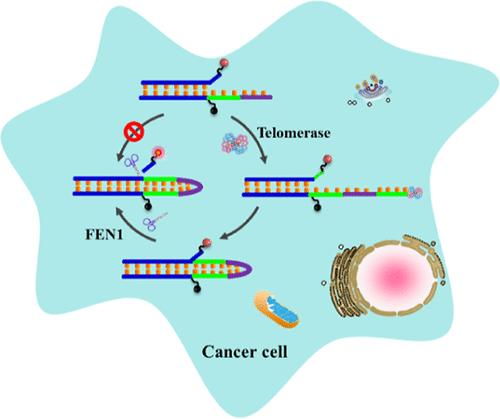
Flap endonuclease 1 (FEN1) is a structure-specific DNA repair enzyme that has emerged as a potential target for cancer diagnosis and treatment. However, existing FEN1 assays often suffer from complicated reaction schemes and laborious procedures, and only a few methods are available for the detection and imaging of FEN1 in living cells. Especially, FEN1 is not exclusive to cancer cells, but it is also shared by normal cells. Consequently, the specific detection of FEN1 in cancer cells remains a challenge. Herein, we develop a simple and selective fluorescent biosensor for the specific imaging of FEN1 in cancer cells and tissues by engineering a FEN1 detection probe with a telomerase-responsive unit. In the presence of telomerase, it induces an extension reaction and subsequent intramolecular reconfiguration of the detection probe, generating a suitable branched DNA structure for FEN1 recognition and facilitating the cleavage of the flap by FEN1 for the recovery of fluorescence signal. Because telomerase is undetectable in normal cells but highly upregulated in cancer cells, the detection probe can only be activated in cancer cells to generate a high signal. This assay is quite simple, with the requirement of merely a single probe for dual enzyme recognition and signal output. With the integration of the single-molecule counting technology, this biosensor can achieve a detection limit of 1.2 × 10⁻⁵ U/μL, and it can accurately detect FEN1 in living cells and clinical tissues, providing a new avenue for FEN1-associated fundamental research and clinical diagnosis.
中文翻译:

内源性端粒酶激活的荧光探针,用于癌细胞和组织中瓣状核酸内切酶 1 的特异性检测和成像
瓣核酸内切酶 1 (FEN1) 是一种结构特异性 DNA 修复酶,已成为癌症诊断和治疗的潜在靶标。然而,现有的 FEN1 检测通常存在复杂的反应方案和费力的程序,并且只有少数方法可用于检测和成像活细胞中的 FEN1。特别是,FEN1 并非癌细胞独有,但它也被正常细胞共享。因此,在癌细胞中特异性检测 FEN1 仍然是一个挑战。在此,我们开发了一种简单的选择性荧光生物传感器,通过设计带有端粒酶反应单元的 FEN1 检测探针,用于癌细胞和组织中 FEN1 的特异性成像。在端粒酶存在下,它诱导检测探针的延伸反应和随后的分子内重配置,产生适合 FEN1 识别的分支 DNA 结构,并促进 FEN1 切割皮瓣以恢复荧光信号。由于端粒酶在正常细胞中检测不到,但在癌细胞中高度上调,因此检测探针只能在癌细胞中激活以产生高信号。该检测非常简单,只需一个探针即可实现双酶识别和信号输出。该生物传感器集成了单分子计数技术,可实现 1.2 × 10⁻⁵ U/μL 的检测限,可以准确检测活细胞和临床组织中的 FEN1,为 FEN1 相关基础研究和临床诊断提供了新的途径。
更新日期:2024-11-20
中文翻译:

内源性端粒酶激活的荧光探针,用于癌细胞和组织中瓣状核酸内切酶 1 的特异性检测和成像
瓣核酸内切酶 1 (FEN1) 是一种结构特异性 DNA 修复酶,已成为癌症诊断和治疗的潜在靶标。然而,现有的 FEN1 检测通常存在复杂的反应方案和费力的程序,并且只有少数方法可用于检测和成像活细胞中的 FEN1。特别是,FEN1 并非癌细胞独有,但它也被正常细胞共享。因此,在癌细胞中特异性检测 FEN1 仍然是一个挑战。在此,我们开发了一种简单的选择性荧光生物传感器,通过设计带有端粒酶反应单元的 FEN1 检测探针,用于癌细胞和组织中 FEN1 的特异性成像。在端粒酶存在下,它诱导检测探针的延伸反应和随后的分子内重配置,产生适合 FEN1 识别的分支 DNA 结构,并促进 FEN1 切割皮瓣以恢复荧光信号。由于端粒酶在正常细胞中检测不到,但在癌细胞中高度上调,因此检测探针只能在癌细胞中激活以产生高信号。该检测非常简单,只需一个探针即可实现双酶识别和信号输出。该生物传感器集成了单分子计数技术,可实现 1.2 × 10⁻⁵ U/μL 的检测限,可以准确检测活细胞和临床组织中的 FEN1,为 FEN1 相关基础研究和临床诊断提供了新的途径。


 京公网安备 11010802027423号