当前位置:
X-MOL 学术
›
Laser Photonics Rev.
›
论文详情
Our official English website, www.x-mol.net, welcomes your
feedback! (Note: you will need to create a separate account there.)
Volumetric Imaging From Raman Perspective: Review and Prospect
Laser & Photonics Reviews ( IF 9.8 ) Pub Date : 2024-11-15 , DOI: 10.1002/lpor.202401444 Nan Wang, Lin Wang, Gong Feng, Maoguo Gong, Weiqi Wang, Shulang Lin, Zhiwei Huang, Xueli Chen
Laser & Photonics Reviews ( IF 9.8 ) Pub Date : 2024-11-15 , DOI: 10.1002/lpor.202401444 Nan Wang, Lin Wang, Gong Feng, Maoguo Gong, Weiqi Wang, Shulang Lin, Zhiwei Huang, Xueli Chen

|
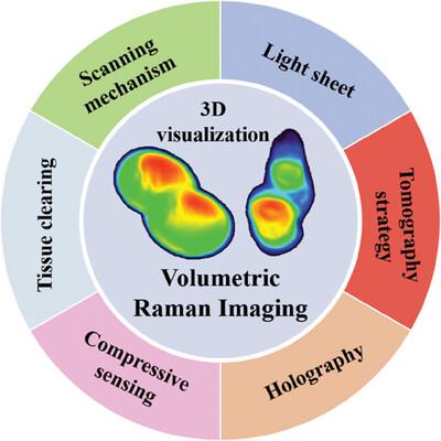
Volumetric imaging, which supports quantitative and comprehensive assessment of a 3D sample from an entire volume, has attracted tremendous attention in biomedical research. Fluorescence imaging techniques, such as optical sectioning and light sheet microscopy, enable to reconstruct the 3D distribution of chemicals within a sample. However, current methods rely on exogenous labels, from which considerable perturbation may be introduced in living systems. Raman imaging offers a feasible solution to visualize components in biological samples in a label‐free manner. Besides, the integration of Raman microscopy with 3D approaches will benefit the research of biomedical samples on novel devices, which is dominated by the strongly enhanced spatial resolution, imaging speed, and overall field of view as well as complemented more details of samples. In this overview, recent achievements in 3D visualization of biological samples from the Raman perspective, are explored including scanning mechanism, light sheet, tomography strategy, compressive sensing, holography, and tissue clearing. Importantly, these platforms are compatible with biomedical research, thus allowing the imaging of chemical constituents and the distribution of samples in a whole volume. As a unique volumetric imaging tool for biological discovery, these methods may provide a strategy to accelerate new discoveries across diverse fields of research.
中文翻译:

从拉曼角度进行体积成像:回顾与展望
体积成像支持对整个体积的 3D 样品进行定量和综合评估,在生物医学研究中引起了极大的关注。荧光成像技术,如光学切片和光片显微镜,能够重建样品中化学物质的 3D 分布。然而,目前的方法依赖于外源标签,从中可能会引入相当大的扰动到生命系统中。拉曼成像提供了一种可行的解决方案,以无标记的方式可视化生物样品中的成分。此外,拉曼显微镜与 3D 方法的整合将有利于在新型设备上进行生物医学样品的研究,其主要优势是大大提高了空间分辨率、成像速度和整体视野,并补充了样品的更多细节。在本概述中,探讨了从拉曼角度对生物样品进行 3D 可视化的最新成就,包括扫描机制、光片、断层扫描策略、压传感、全息术和组织透明化。重要的是,这些平台与生物医学研究兼容,因此可以对化学成分进行成像和整个体积的样品分布。作为用于生物发现的独特体积成像工具,这些方法可能提供一种策略来加速不同研究领域的新发现。
更新日期:2024-11-15
中文翻译:

从拉曼角度进行体积成像:回顾与展望
体积成像支持对整个体积的 3D 样品进行定量和综合评估,在生物医学研究中引起了极大的关注。荧光成像技术,如光学切片和光片显微镜,能够重建样品中化学物质的 3D 分布。然而,目前的方法依赖于外源标签,从中可能会引入相当大的扰动到生命系统中。拉曼成像提供了一种可行的解决方案,以无标记的方式可视化生物样品中的成分。此外,拉曼显微镜与 3D 方法的整合将有利于在新型设备上进行生物医学样品的研究,其主要优势是大大提高了空间分辨率、成像速度和整体视野,并补充了样品的更多细节。在本概述中,探讨了从拉曼角度对生物样品进行 3D 可视化的最新成就,包括扫描机制、光片、断层扫描策略、压传感、全息术和组织透明化。重要的是,这些平台与生物医学研究兼容,因此可以对化学成分进行成像和整个体积的样品分布。作为用于生物发现的独特体积成像工具,这些方法可能提供一种策略来加速不同研究领域的新发现。


 京公网安备 11010802027423号