当前位置:
X-MOL 学术
›
Ind. Crops Prod.
›
论文详情
Our official English website, www.x-mol.net, welcomes your
feedback! (Note: you will need to create a separate account there.)
Microscopic spatiotemporal changes in cell wall cellulose and pectin during Nicotiana tabacum L. leaf growth and senescence based on label-free Raman microspectroscopic imaging combined with multivariate curve resolution
Industrial Crops and Products ( IF 5.6 ) Pub Date : 2024-10-19 , DOI: 10.1016/j.indcrop.2024.119865 Mei Li, Ke-Su Wei, Yuan Xue, Sheng-Jiang Wu, Ya-Juan Liu, Dong-Mei Chen, Xiu-Fang Yan, Chao Kang
Industrial Crops and Products ( IF 5.6 ) Pub Date : 2024-10-19 , DOI: 10.1016/j.indcrop.2024.119865 Mei Li, Ke-Su Wei, Yuan Xue, Sheng-Jiang Wu, Ya-Juan Liu, Dong-Mei Chen, Xiu-Fang Yan, Chao Kang

|
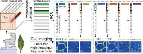
The plant cell wall, composed mainly of polysaccharides, lignin, and structural proteins, supports the architecture, mechanics, and functions of plants. Developing appropriate chemical imaging methods to study spatiotemporal changes of cell wall structural components at the microscopic level is important for understanding plant growth and senescence. In this study, tobacco (Nicotiana tabacum L.), a widely cultivated economic crop and model plant, was selected as the research object. Based on Raman confocal imaging combined with a multivariate curve resolution model, a label-free, in situ, high-throughput and high specificity imaging method for cellulose, high methylated pectin, and low methylated pectin in tobacco leaf cell wall was established to study their microscopic spatiotemporal changes during leaf growth and senescence (flue-curing) processes. The results based on the proposed method revealed that cellulose and pectin levels in the midrib cell wall gradually increased as the leaves matured, from appeared mainly at the cell corners and middle lamella respectively, to appeared in the cell corners, middle lamella, and cell wall. The same trend was observed in the lateral vein cell walls, where cellulose and pectin levels gradually increased. During the flue-curing process, cellulose and highly methylated pectin degraded. The proposed chemical imaging method is expected to provide a label-free, in situ, and high-throughput cell imaging technique for investigating the microscopic spatiotemporal distribution of the main structural components of the leaf cell wall.
中文翻译:

基于无标记拉曼显微成像结合多变量曲线分辨率的烟草叶片生长和衰老过程中细胞壁纤维素和果胶的微观时空变化
植物细胞壁主要由多糖、木质素和结构蛋白组成,支持植物的结构、力学和功能。开发适当的化学成像方法以在微观水平上研究细胞壁结构成分的时空变化对于理解植物生长和衰老非常重要。本研究以广泛栽培的经济作物和模式植物烟草 (Nicotiana tabacum L.) 为研究对象。基于拉曼共聚焦成像结合多元曲线分辨率模型,建立了烟草叶壁中纤维素、高甲基化果胶和低甲基化果胶的无标记、原位、高通量、高特异性成像方法,研究它们在叶片生长和衰老(烤烟)过程中的微观时空变化。基于该方法的结果表明,随着叶片的成熟,中脉细胞壁中的纤维素和果胶水平逐渐增加,从主要出现在细胞角和中间薄片,发展到出现在细胞角、中间薄片和细胞壁。在侧脉细胞壁中观察到相同的趋势,其中纤维素和果胶水平逐渐增加。在烤烟固化过程中,纤维素和高度甲基化的果胶降解。所提出的化学成像方法有望提供一种无标记、原位和高通量细胞成像技术,用于研究叶细胞壁主要结构成分的微观时空分布。
更新日期:2024-10-20
中文翻译:

基于无标记拉曼显微成像结合多变量曲线分辨率的烟草叶片生长和衰老过程中细胞壁纤维素和果胶的微观时空变化
植物细胞壁主要由多糖、木质素和结构蛋白组成,支持植物的结构、力学和功能。开发适当的化学成像方法以在微观水平上研究细胞壁结构成分的时空变化对于理解植物生长和衰老非常重要。本研究以广泛栽培的经济作物和模式植物烟草 (Nicotiana tabacum L.) 为研究对象。基于拉曼共聚焦成像结合多元曲线分辨率模型,建立了烟草叶壁中纤维素、高甲基化果胶和低甲基化果胶的无标记、原位、高通量、高特异性成像方法,研究它们在叶片生长和衰老(烤烟)过程中的微观时空变化。基于该方法的结果表明,随着叶片的成熟,中脉细胞壁中的纤维素和果胶水平逐渐增加,从主要出现在细胞角和中间薄片,发展到出现在细胞角、中间薄片和细胞壁。在侧脉细胞壁中观察到相同的趋势,其中纤维素和果胶水平逐渐增加。在烤烟固化过程中,纤维素和高度甲基化的果胶降解。所提出的化学成像方法有望提供一种无标记、原位和高通量细胞成像技术,用于研究叶细胞壁主要结构成分的微观时空分布。


 京公网安备 11010802027423号