Molecular Psychiatry ( IF 9.6 ) Pub Date : 2024-10-16 , DOI: 10.1038/s41380-024-02781-5 Daniel S. Scott, Muthumeenakshi Subramanian, Jun Yamamoto, Carol A. Tamminga
|
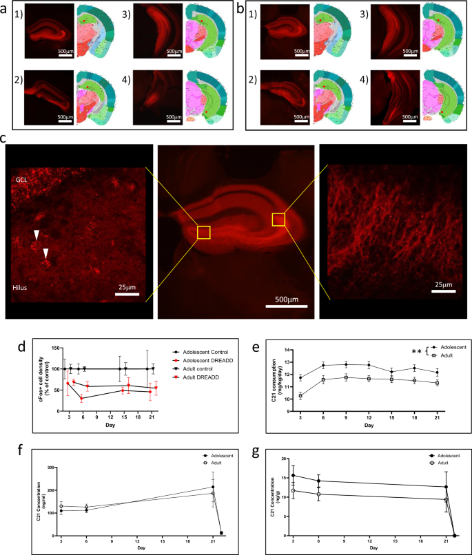
Decades of research into the function of the medial temporal lobe has driven curiosity around clinical outcomes associated with hippocampal dysfunction, including psychosis. Post-mortem analyses of brain tissue from human schizophrenia brain show decreased expression of the NMDAR subunit GluN1 confined to the dentate gyrus with evidence of downstream hippocampal hyperactivity in CA3 and CA1. Little is known about the mechanisms of the emergence of hippocampal hyperactivity as a putative psychosis biomarker. We have developed a reverse-translation mouse to study critical neural features. We had previously studied a dentate gyrus (DG)-specific GluN1 KO, which displays hippocampal hyperactivity and a psychosis-relevant behavioral phenotype. Here, we expressed an inhibitory DREADD (pAAV-CaMKIIa-hM4D(Gi)-mCherry) in granule cells of the mouse dentate gyrus, and continuously inhibited the region for 21 days in adolescent (6 weeks) and adult (10 weeks) C57BL/6 J mice with DREADD agonist Compound 21 (C21). Following this period, we quantified activity in the hippocampal subfields by assessing cFos expression, hippocampally mediated behaviors, and hippocampal local field potential with an intracerebral probe with continual monitoring over time. DG inhibition during adolescence generates an increase in hippocampal activity in CA3 and CA1, impairs social cognition and spatial working memory, as well as shows evidence of increased activity in local field potentials as spontaneous synchronous bursts of activity, which we term hyper-synchronous events (HSEs) in hippocampus. The same DG inhibition delivered during adulthood in the mouse lacks these outcomes. These results suggest a sensitive period in development in which the hippocampus is susceptible to DG inhibition resulting in hippocampal hyperactivity and psychosis-like behavioral outcomes.
中文翻译:
精神分裂症病理逆向翻译成小鼠显示海马多动、精神病行为和超同步事件
数十年来对内侧颞叶功能的研究引发了人们对与海马功能障碍(包括精神病)相关的临床结果的好奇心。对人类精神分裂症大脑脑组织的尸检分析显示,局限于齿状回的 NMDAR 亚基 GluN1 表达降低,有证据表明 CA3 和 CA1 下游海马多动。关于海马多动症作为推定的精神病生物标志物出现的机制知之甚少。我们开发了一种反向翻译小鼠来研究关键神经特征。我们之前研究了齿状回 (DG) 特异性 GluN1 KO,它显示海马多动和精神病相关行为表型。在这里,我们在小鼠齿状回的颗粒细胞中表达抑制性 DREADD (pAAV-CaMKIIa-hM4D(Gi)-mCherry),并在青少年 (6 周) 和成年 (10 周) C57BL/6 J 小鼠中连续抑制该区域 21 天DREADD 激动剂化合物 21 (C21)。在此期间,我们通过评估 cFos 表达、海马介导的行为和海马局部场电位来量化海马子场中的活动,并使用脑内探针进行持续监测。青春期的 DG 抑制导致 CA3 和 CA1 海马活动增加,损害社会认知和空间工作记忆,并显示局部场电位活动增加的证据,表现为自发的同步活动爆发,我们称之为海马体中的超同步事件 (HSE)。在小鼠成年期提供的相同 DG 抑制缺乏这些结果。 这些结果表明,海马体在发育过程中容易受到 DG 抑制,导致海马体多动和精神病样行为结果。


 京公网安备 11010802027423号