当前位置:
X-MOL 学术
›
Int. J. Plasticity
›
论文详情
Our official English website, www.x-mol.net, welcomes your
feedback! (Note: you will need to create a separate account there.)
3D strain heterogeneity and fracture studied by X-ray tomography and crystal plasticity in an aluminium alloy
International Journal of Plasticity ( IF 9.4 ) Pub Date : 2024-10-12 , DOI: 10.1016/j.ijplas.2024.104146 Maryse Gille, Henry Proudhon, Jette Oddershede, Romain Quey, Thilo F. Morgeneyer
International Journal of Plasticity ( IF 9.4 ) Pub Date : 2024-10-12 , DOI: 10.1016/j.ijplas.2024.104146 Maryse Gille, Henry Proudhon, Jette Oddershede, Romain Quey, Thilo F. Morgeneyer

|
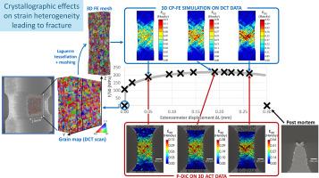
Strong correlations between measured strain fields and 3D crystal plasticity finite element (CP-FE) predictions based on the real microstructure are found for a plane strain tensile specimen made of 6016 T4 aluminium alloy. This is achieved using multimodal X-ray lab tomography giving access to both the initial grain structure and the strain evolution. The real microstructure of the central region of interest (ROI) of the undeformed specimen is obtained non destructively using lab-based diffraction contrast tomography (DCT) and meshing. An in situ tensile test, using absorption contrast tomography (ACT) is then performed for twelve loading increments up to fracture. Taking advantage of the plane strain condition, the evolution of the internal strain field is measured by two-dimensional digital image correlation (DIC) in the material bulk using the natural speckle provided by intermetallic particles. Early strain heterogeneities in the form of slanted bands, that are spatially stable over time, are revealed and the fracture path – determined from the post mortem scan – is found to coincide with the bands exhibiting maximum strain. CP-FE simulations are performed on the meshed microstructure of the specimen acquired by DCT and are compared with image correlation measurements. The measured strain fields are well described by 3D CP-FE predictions, whilst it is shown that neither a macroscopic anisotropic plasticity model nor a CP-FE simulation with random grain orientations could reproduce the measurements.
中文翻译:

通过 X 射线断层扫描和晶体塑性研究铝合金的三维应变异质性和断裂
对于 6016 T4 铝合金制成的平面应变拉伸试样,发现测得的应变场与基于真实微观结构的 3D 晶体塑性有限元 (CP-FE) 预测之间存在很强的相关性。这是使用多模态 X 射线实验室断层扫描实现的,可以同时获得初始晶粒结构和应变演变。使用基于实验室的衍射对比断层扫描 (DCT) 和网格划分,以非破坏性方式获得未变形样品的中心感兴趣区域 (ROI) 的真实微观结构。然后使用吸收对比断层扫描 (ACT) 进行原位拉伸试验,进行 12 次载荷增量,直至骨折。利用平面应变条件,利用金属间化合物颗粒提供的自然散斑,通过材料块体中的二维数字图像相关 (DIC) 来测量内部应变场的演变。揭示了倾斜带形式的早期应变异质性,这些异常性在空间上随时间保持稳定,并且发现断裂路径 - 通过尸检扫描确定 - 与表现出最大应变的条带重合。对 DCT 采集的样品的网格微观结构进行 CP-FE 仿真,并与图像相关测量进行比较。3D CP-FE 预测很好地描述了测得的应变场,同时表明宏观各向异性塑性模型和具有随机晶粒取向的 CP-FE 模拟都无法重现测量结果。
更新日期:2024-10-12
中文翻译:

通过 X 射线断层扫描和晶体塑性研究铝合金的三维应变异质性和断裂
对于 6016 T4 铝合金制成的平面应变拉伸试样,发现测得的应变场与基于真实微观结构的 3D 晶体塑性有限元 (CP-FE) 预测之间存在很强的相关性。这是使用多模态 X 射线实验室断层扫描实现的,可以同时获得初始晶粒结构和应变演变。使用基于实验室的衍射对比断层扫描 (DCT) 和网格划分,以非破坏性方式获得未变形样品的中心感兴趣区域 (ROI) 的真实微观结构。然后使用吸收对比断层扫描 (ACT) 进行原位拉伸试验,进行 12 次载荷增量,直至骨折。利用平面应变条件,利用金属间化合物颗粒提供的自然散斑,通过材料块体中的二维数字图像相关 (DIC) 来测量内部应变场的演变。揭示了倾斜带形式的早期应变异质性,这些异常性在空间上随时间保持稳定,并且发现断裂路径 - 通过尸检扫描确定 - 与表现出最大应变的条带重合。对 DCT 采集的样品的网格微观结构进行 CP-FE 仿真,并与图像相关测量进行比较。3D CP-FE 预测很好地描述了测得的应变场,同时表明宏观各向异性塑性模型和具有随机晶粒取向的 CP-FE 模拟都无法重现测量结果。


 京公网安备 11010802027423号