Light: Science & Applications ( IF 20.6 ) Pub Date : 2024-10-11 , DOI: 10.1038/s41377-024-01619-7 Patrick C. Chaumet, Pierre Bon, Guillaume Maire, Anne Sentenac, Guillaume Baffou
|
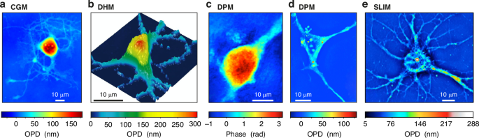
Quantitative phase microscopies (QPMs) play a pivotal role in bio-imaging, offering unique insights that complement fluorescence imaging. They provide essential data on mass distribution and transport, inaccessible to fluorescence techniques. Additionally, QPMs are label-free, eliminating concerns of photobleaching and phototoxicity. However, navigating through the array of available QPM techniques can be complex, making it challenging to select the most suitable one for a particular application. This tutorial review presents a thorough comparison of the main QPM techniques, focusing on their accuracy in terms of measurement precision and trueness. We focus on 8 techniques, namely digital holographic microscopy (DHM), cross-grating wavefront microscopy (CGM), which is based on QLSI (quadriwave lateral shearing interferometry), diffraction phase microscopy (DPM), differential phase-contrast (DPC) microscopy, phase-shifting interferometry (PSI) imaging, Fourier phase microscopy (FPM), spatial light interference microscopy (SLIM), and transport-of-intensity equation (TIE) imaging. For this purpose, we used a home-made numerical toolbox based on discrete dipole approximation (IF-DDA). This toolbox is designed to compute the electromagnetic field at the sample plane of a microscope, irrespective of the object’s complexity or the illumination conditions. We upgraded this toolbox to enable it to model any type of QPM, and to take into account shot noise. In a nutshell, the results show that DHM and PSI are inherently free from artefacts and rather suffer from coherent noise; In CGM, DPC, DPM and TIE, there is a trade-off between precision and trueness, which can be balanced by varying one experimental parameter; FPM and SLIM suffer from inherent artefacts that cannot be discarded experimentally in most cases, making the techniques not quantitative especially for large objects covering a large part of the field of view, such as eukaryotic cells.
中文翻译:
定量相显微镜:准确性比较
定量相显微镜 (QPM) 在生物成像中起着关键作用,为荧光成像提供了独特的见解。它们提供了荧光技术无法获得的有关质量分布和传输的基本数据。此外,QPM 是无标记的,无需担心光漂白和光毒性。然而,在一系列可用的 QPM 技术中导航可能很复杂,因此很难为特定应用选择最合适的技术。本教学案例综述对主要的 QPM 技术进行了全面比较,重点介绍了它们在测量精度和真实度方面的准确性。我们专注于 8 种技术,即数字全息显微镜 (DHM)、基于 QLSI(四波横向剪切干涉法)的交叉光栅波前显微镜 (CGM)、衍射相位显微镜 (DPM)、差分相差 (DPC) 显微镜、相移干涉法 (PSI) 成像、傅里叶相位显微镜 (FPM)、空间光干涉显微镜 (SLIM) 和强度传输方程 (TIE) 成像。为此,我们使用了一个基于离散偶极子近似 (IF-DDA) 的自制数值工具箱。该工具箱旨在计算显微镜样品平面上的电磁场,而与物体的复杂程度或照明条件无关。我们升级了这个工具箱,使其能够对任何类型的 QPM 进行建模,并考虑散粒噪声。 简而言之,结果表明 DHM 和 PSI 本质上没有伪影,而是受到相干噪声的影响;在 CGM、DPC、DPM 和 TIE 中,精度和真实度之间存在权衡,这可以通过改变一个实验参数来平衡;FPM 和 SLIM 存在固有的伪影,在大多数情况下无法通过实验丢弃,这使得这些技术不是定量的,特别是对于覆盖大部分视野的大型物体,例如真核细胞。


 京公网安备 11010802027423号