European Journal of Nuclear Medicine and Molecular Imaging ( IF 8.6 ) Pub Date : 2024-10-09 , DOI: 10.1007/s00259-024-06938-w Benjamin F. Combes, Sandeep Kumar Kalva, Pierre-Louis Benveniste, Agathe Tournant, Man Hoi Law, Joshua Newton, Maik Krüger, Rebecca Z. Weber, Inês Dias, Daniela Noain, Xose Luis Dean-Ben, Uwe Konietzko, Christian R. Baumann, Per-Göran Gillberg, Christoph Hock, Roger M. Nitsch, Julien Cohen-Adad, Daniel Razansky, Ruiqing Ni
|
Purpose
Metabolism and bioenergetics in the central nervous system play important roles in the pathophysiology of Parkinson’s disease (PD). Here, we employed a multimodal imaging approach to assess oxygenation changes in the spinal cord of the transgenic M83 murine model of PD overexpressing the mutated A53T alpha-synuclein form in comparison with non-transgenic littermates.
Methods
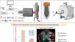
In vivo spiral volumetric optoacoustic tomography (SVOT) was performed to assess oxygen saturation (sO2) in the spinal cords of M83 mice and non-transgenic littermates. Ex vivo high-field T1-weighted (T1w) magnetic resonance imaging (MRI) at 9.4T was used to assess volumetric alterations in the spinal cord. 3D SVOT analysis and deep learning-based automatic segmentation of T1w MRI data for the mouse spinal cord were developed for quantification. Immunostaining for phosphorylated alpha-synuclein (pS129 α-syn), as well as vascular organization (CD31 and GLUT1), was performed after MRI scan.
Results
In vivo SVOT imaging revealed a lower sO2SVOT in the spinal cord of M83 mice compared to non-transgenic littermates at sub-100 μm spatial resolution. Ex vivo MRI-assisted by in-house developed deep learning-based automatic segmentation (validated by manual analysis) revealed no volumetric atrophy in the spinal cord of M83 mice compared to non-transgenic littermates at 50 μm spatial resolution. The vascular network was not impaired in the spinal cord of M83 mice in the presence of pS129 α-syn accumulation.
Conclusion
We developed tools for deep-learning-based analysis for the segmentation of mouse spinal cord structural MRI data, and volumetric analysis of sO2SVOT data. We demonstrated non-invasive high-resolution imaging of reduced sO2SVOT in the absence of volumetric structural changes in the spinal cord of PD M83 mouse model.
中文翻译:
帕金森病 M83 小鼠脊髓氧饱和度降低的螺旋体积光声断层扫描
目的
中枢神经系统的新陈代谢和生物能量学在帕金森病 (PD) 的病理生理学中起重要作用。在这里,我们采用多模态成像方法来评估与非转基因同窝同窝小鼠相比,过表达突变的 A53T α-突触核蛋白形式的 PD 的转基因 M83 小鼠模型脊髓的氧合变化。
方法
进行体内螺旋体积光声断层扫描 (SVOT) 以评估 M83 小鼠和非转基因同窝小鼠脊髓中的氧饱和度 (sO2)。使用 9.4T 的离体高场 T1 加权 (T1w) 磁共振成像 (MRI) 评估脊髓的体积变化。开发了 3D SVOT 分析和基于深度学习的小鼠脊髓 T1w MRI 数据自动分割用于量化。MRI 扫描后对磷酸化 α-突触核蛋白 (pS129 α-syn) 以及血管组织 (CD31 和 GLUT1) 进行免疫染色。
结果
体内 SVOT 成像显示,在亚 100 μm 空间分辨率下,与非转基因同窝小鼠相比,M83 小鼠脊髓中的 sO2SVOT 较低。由内部开发的基于深度学习的自动分割(通过人工分析验证)辅助的离体 MRI 显示,在 50 μm 空间分辨率下,与非转基因同窝小鼠相比,M83 小鼠的脊髓没有体积萎缩。在 pS129 α-syn 积累存在的情况下,M83 小鼠脊髓中的血管网络未受损。
结论
我们开发了基于深度学习的分析工具,用于小鼠脊髓结构 MRI 数据的分割和 sO2SVOT 数据的体积分析。我们展示了在 PD M83 小鼠模型的脊髓没有体积结构变化的情况下,sO2SVOT 降低的无创高分辨率成像。


 京公网安备 11010802027423号