Our official English website, www.x-mol.net, welcomes your
feedback! (Note: you will need to create a separate account there.)
Rapid low-cost assembly of modular microvessel-on-a-chip with benchtop xurography
Lab on a Chip ( IF 6.1 ) Pub Date : 2024-10-07 , DOI: 10.1039/d4lc00565a Shashwat S. Agarwal, Marcos Cortes-Medina, Jacob C. Holter, Alex Avendano, Joseph W. Tinapple, Joseph M. Barlage, Miles M. Menyhert, Lotanna M. Onua, Jonathan W. Song
Lab on a Chip ( IF 6.1 ) Pub Date : 2024-10-07 , DOI: 10.1039/d4lc00565a Shashwat S. Agarwal, Marcos Cortes-Medina, Jacob C. Holter, Alex Avendano, Joseph W. Tinapple, Joseph M. Barlage, Miles M. Menyhert, Lotanna M. Onua, Jonathan W. Song

|
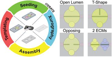
Blood and lymphatic vessels in the body are central to molecular and cellular transport, tissue repair, and pathophysiology. Several approaches have been employed for engineering microfabricated blood and lymphatic vessels in vitro, yet traditionally these approaches require specialized equipment, facilities, and research training beyond the capabilities of many biomedical laboratories. Here we present xurography as an inexpensive, accessible, and versatile rapid prototyping technique for engineering cylindrical and lumenized microvessels. Using a benchtop xurographer, or a cutting plotter, we fabricated modular multi-layer poly(dimethylsiloxane) (PDMS)-based microphysiological systems (MPS) that house endothelial-lined microvessels approximately 260 μm in diameter embedded within a user-defined 3-D extracellular matrix (ECM). We validated the vascularized MPS (or vessel-on-a-chip) by quantifying changes in blood vessel permeability due to the pro-angiogenic chemokine CXCL12. Moreover, we demonstrated the reconfigurable versatility of this approach by engineering a total of four distinct vessel-ECM arrangements, which were obtained by only minor adjustments to a few steps of the fabrication process. Several of these arrangements, such as ones that incorporate close-ended vessel structures and spatially distinct ECM compartments along the same microvessel, have not been widely achieved with other microfabrication strategies. Therefore, we anticipate that our low-cost and easy-to-implement fabrication approach will facilitate broader adoption of MPS with customizable vascular architectures and ECM components while reducing the turnaround time required for iterative designs.
中文翻译:

使用台式 Xurography 快速低成本组装模块化芯片微容器
体内的血液和淋巴管是分子和细胞运输、组织修复和病理生理学的核心。已经采用了几种方法在体外设计微加工血液和淋巴管,但传统上这些方法需要专门的设备、设施和研究培训,超出了许多生物医学实验室的能力。在这里,我们将 Xurography 介绍为一种廉价、可访问且用途广泛的快速原型技术,用于设计圆柱形和管腔化微血管。使用台式超声技师或刻字机,我们制造了模块化多层基于聚(二甲基硅氧烷)(PDMS)的微生理系统 (MPS),该系统包含直径约 260 μm 的内皮衬里微血管,嵌入用户定义的 3-D 细胞外基质 (ECM)。我们通过量化促血管生成趋化因子 CXCL12 引起的血管通透性变化来验证血管化 MPS(或芯片上血管)。此外,我们通过设计总共四种不同的容器-ECM 布置来证明这种方法的可重构多功能性,这些排列只需对制造过程的几个步骤进行微小调整即可获得。其中一些布置,例如沿同一微血管结合闭端血管结构和空间上不同的 ECM 隔室的布置,尚未通过其他微加工策略广泛实现。因此,我们预计我们的低成本且易于实施的制造方法将促进 MPS 的广泛采用,同时减少迭代设计所需的周转时间。
更新日期:2024-10-07
中文翻译:

使用台式 Xurography 快速低成本组装模块化芯片微容器
体内的血液和淋巴管是分子和细胞运输、组织修复和病理生理学的核心。已经采用了几种方法在体外设计微加工血液和淋巴管,但传统上这些方法需要专门的设备、设施和研究培训,超出了许多生物医学实验室的能力。在这里,我们将 Xurography 介绍为一种廉价、可访问且用途广泛的快速原型技术,用于设计圆柱形和管腔化微血管。使用台式超声技师或刻字机,我们制造了模块化多层基于聚(二甲基硅氧烷)(PDMS)的微生理系统 (MPS),该系统包含直径约 260 μm 的内皮衬里微血管,嵌入用户定义的 3-D 细胞外基质 (ECM)。我们通过量化促血管生成趋化因子 CXCL12 引起的血管通透性变化来验证血管化 MPS(或芯片上血管)。此外,我们通过设计总共四种不同的容器-ECM 布置来证明这种方法的可重构多功能性,这些排列只需对制造过程的几个步骤进行微小调整即可获得。其中一些布置,例如沿同一微血管结合闭端血管结构和空间上不同的 ECM 隔室的布置,尚未通过其他微加工策略广泛实现。因此,我们预计我们的低成本且易于实施的制造方法将促进 MPS 的广泛采用,同时减少迭代设计所需的周转时间。


 京公网安备 11010802027423号