Our official English website, www.x-mol.net, welcomes your
feedback! (Note: you will need to create a separate account there.)
Cathodoluminescent and Characteristic X-Ray-Emissive Rare-Earth-Doped Core/Shell Protein Labels for Spectromicroscopic Analysis of Cell Surface Receptors
Small ( IF 13.0 ) Pub Date : 2024-09-09 , DOI: 10.1002/smll.202404309 Sebastian Habermann 1, 2 , Lukas R H Gerken 1, 2 , Mathieu Kociak 3 , Christian Monachon 4 , Vera M Kissling 2 , Alexander Gogos 1, 2 , Inge K Herrmann 1, 2, 5, 6
Small ( IF 13.0 ) Pub Date : 2024-09-09 , DOI: 10.1002/smll.202404309 Sebastian Habermann 1, 2 , Lukas R H Gerken 1, 2 , Mathieu Kociak 3 , Christian Monachon 4 , Vera M Kissling 2 , Alexander Gogos 1, 2 , Inge K Herrmann 1, 2, 5, 6
Affiliation

|
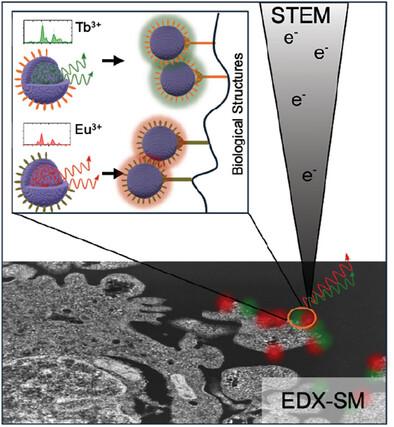
Understanding the localization and the interactions of biomolecules at the nanoscale and in the cellular context remains challenging. Electron microscopy (EM), unlike light-based microscopy, gives access to the cellular ultrastructure yet results in grey-scale images and averts unambiguous (co-)localization of biomolecules. Multimodal nanoparticle-based protein labels for correlative cathodoluminescence electron microscopy (CCLEM) and energy-dispersive X-ray spectromicroscopy (EDX-SM) are presented. The single-particle STEM-cathodoluminescence (CL) and characteristic X-ray emissivity of sub-20 nm lanthanide-doped nanoparticles are exploited as unique spectral fingerprints for precise label localization and identification. To maximize the nanoparticle brightness, lanthanides are incorporated in a low-phonon host lattice and separated from the environment using a passivating shell. The core/shell nanoparticles are then functionalized with either folic (terbium-doped) or caffeic acid (europium-doped). Their potential for (protein-)labeling is successfully demonstrated using HeLa cells expressing different surface receptors that bind to folic or caffeic acid, respectively. Both particle populations show single-particle CL emission along with a distinctive energy-dispersive X-ray signal, with the latter enabling color-based localization of receptors within swift imaging times well below 2 min per
中文翻译:

阴极发光和特征 X 射线发射稀土掺杂核/壳蛋白标记,用于细胞表面受体的光谱显微镜分析
了解生物分子在纳米尺度和细胞环境中的定位和相互作用仍然具有挑战性。与基于光学的显微镜不同,电子显微镜 (EM) 可以访问细胞超微结构,但会产生灰度图像,并避免了生物分子的明确(共)定位。介绍了用于相关阴极发光电子显微镜 (CCLEM) 和能量色散 X 射线光谱显微镜 (EDX-SM) 的基于多模式纳米粒子的蛋白质标记。利用单粒子 STEM 阴极发光 (CL) 和亚 20 nm 镧系元素掺杂纳米颗粒的特征 X 射线发射率作为独特的光谱指纹,用于精确的标记定位和识别。为了最大限度地提高纳米颗粒的亮度,镧系元素被掺入低声子主晶格中,并使用钝化壳层与环境分离。然后用叶酸(掺铽)或咖啡酸(掺铕)对核/壳纳米颗粒进行功能化。使用分别表达与叶酸或咖啡酸结合的不同表面受体的 HeLa 细胞成功证明了它们的(蛋白质)标记潜力。两种粒子群都显示单粒子 CL 发射以及独特的能量色散 X 射线信号,后者能够在远低于每
更新日期:2024-09-09
中文翻译:

阴极发光和特征 X 射线发射稀土掺杂核/壳蛋白标记,用于细胞表面受体的光谱显微镜分析
了解生物分子在纳米尺度和细胞环境中的定位和相互作用仍然具有挑战性。与基于光学的显微镜不同,电子显微镜 (EM) 可以访问细胞超微结构,但会产生灰度图像,并避免了生物分子的明确(共)定位。介绍了用于相关阴极发光电子显微镜 (CCLEM) 和能量色散 X 射线光谱显微镜 (EDX-SM) 的基于多模式纳米粒子的蛋白质标记。利用单粒子 STEM 阴极发光 (CL) 和亚 20 nm 镧系元素掺杂纳米颗粒的特征 X 射线发射率作为独特的光谱指纹,用于精确的标记定位和识别。为了最大限度地提高纳米颗粒的亮度,镧系元素被掺入低声子主晶格中,并使用钝化壳层与环境分离。然后用叶酸(掺铽)或咖啡酸(掺铕)对核/壳纳米颗粒进行功能化。使用分别表达与叶酸或咖啡酸结合的不同表面受体的 HeLa 细胞成功证明了它们的(蛋白质)标记潜力。两种粒子群都显示单粒子 CL 发射以及独特的能量色散 X 射线信号,后者能够在远低于每


 京公网安备 11010802027423号