Virchows Archiv ( IF 3.4 ) Pub Date : 2024-09-03 , DOI: 10.1007/s00428-024-03903-8 Takumi Kitaoka 1, 2 , Kenji Harada 3, 4 , Shingo Sakashita 3 , Motohiro Kojima 3 , Tetsuro Taki 1 , Takeshi Kuwata 5 , Takahiro Kinoshita 6 , Mitsuru Futakuchi 2 , Genichiro Ishii 1, 7 , Naoya Sakamoto 3
|
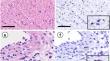
Gremlin 1 (GREM1) is an antagonist of bone morphogenetic protein (BMP). GREM1 is expressed in the stromal cells of various carcinomas and promotes tumor progression by suppressing BMP signaling. We designed this study to establish an evaluation strategy for GREM1 expression, focusing on the tumor stroma, and to examine its clinicopathological significance in gastric cancer (GC) progression. We employed RNA in situ hybridization (ISH) to evaluate the prognostic value of GREM1 expression in a cohort of 104 surgically resected GC cases and assessed ISH scores according to previous reports. GREM1 expression was observed in tumor stromal cells, including fibroblasts. We defined GREM1-positive cells as those expressing ISH score ≥ 3 and quantified the number of GREM1-positive cells using image analysis software. We examined the relationship between the number of GREM1-positive cells in the tumor stroma and clinicopathological features. The number of GREM1-positive cells per tumor stroma ranged from 0 to 714.7 cells/mm2 (median, 1.65 cells/mm2). We divided the 104 GC cases into GREM1-High and GREM1-Low expression groups based on the abovementioned median value. GREM1-High expression group was significantly associated with a more advanced pT grade, pN grade, lymphatic invasion, and venous invasion. Kaplan–Meier analysis showed significantly poorer survival in the GREM1-High expression group than in the GREM1-Low expression group. These results indicated that GREM1 expression in GC is localized in tumor stromal cells, and that high GREM1 expression in the tumor stroma could be a poor prognostic factor.
中文翻译:
使用全玻片成像对整个肿瘤基质中的 Gremlin 1 进行定量及其在胃癌中的临床病理意义
Gremlin 1 (GREM1) 是骨形态发生蛋白 (BMP) 的拮抗剂。 GREM1 在多种癌症的基质细胞中表达,并通过抑制 BMP 信号传导促进肿瘤进展。我们设计本研究是为了建立GREM1表达的评估策略,重点关注肿瘤基质,并检查其在胃癌 (GC) 进展中的临床病理学意义。我们采用 RNA 原位杂交 (ISH) 来评估 104 例手术切除的 GC 病例中GREM1表达的预后价值,并根据以前的报告评估 ISH 评分。在肿瘤基质细胞(包括成纤维细胞)中观察到GREM1表达。我们将GREM1阳性细胞定义为表达 ISH 评分≥ 3 的细胞,并使用图像分析软件量化GREM1阳性细胞的数量。我们检查了肿瘤基质中GREM1阳性细胞的数量与临床病理特征之间的关系。每个肿瘤基质的GREM1阳性细胞数量范围为0至714.7个细胞/mm 2 (中位数,1.65个细胞/mm 2 )。我们根据上述中值将104例GC病例分为GREM1高表达组和GREM1低表达组。 GREM1高表达组与更晚期的pT分级、pN分级、淋巴侵犯和静脉侵犯显着相关。 Kaplan-Meier 分析显示, GREM1高表达组的生存率明显低于GREM1低表达组。 这些结果表明,GC 中的GREM1表达位于肿瘤基质细胞中,肿瘤基质中的高GREM1表达可能是一个不良的预后因素。


 京公网安备 11010802027423号