当前位置:
X-MOL 学术
›
Phys. Chem. Chem. Phys.
›
论文详情
Our official English website, www.x-mol.net, welcomes your
feedback! (Note: you will need to create a separate account there.)
Metal–ligand delocalization of iron and cobalt porphyrin complexes in aqueous solutions probed by soft X-ray absorption spectroscopy
Physical Chemistry Chemical Physics ( IF 2.9 ) Pub Date : 2024-08-27 , DOI: 10.1039/d4cp02140a Masanari Nagasaka 1, 2 , Shota Tsuru 3, 4 , Yasuyuki Yamada 5, 6
Physical Chemistry Chemical Physics ( IF 2.9 ) Pub Date : 2024-08-27 , DOI: 10.1039/d4cp02140a Masanari Nagasaka 1, 2 , Shota Tsuru 3, 4 , Yasuyuki Yamada 5, 6
Affiliation

|
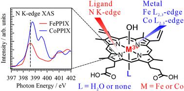
Metal–ligand delocalization of metal porphyrin complexes in aqueous solutions was investigated by analyzing the electronic structure of both the metal and ligand sides using soft X-ray absorption spectroscopy (XAS) at the metal L2,3-edges and nitrogen K-edge, respectively. In the N K-edge XAS spectra of the ligands, the energies of the C![[double bond, length as m-dash]](https://www.rsc.org/images/entities/char_e001.gif) N π* peaks of cobalt protoporphyrin IX (CoPPIX) are higher than iron protoporphyrin IX (FePPIX). The energy difference between the two lowest peaks in the XAS spectrum of CoPPIX is also larger than that of FePPIX. Nitrogen K-edge inner-shell calculations of metalloporphyrins with different central metals indicate that the energy differences between these peaks reflect the electronic configurations and spin multiplicities of metalloporphyrins. We also investigated the hydration structure of CoPPIX in aqueous solution by analyzing the electronic structure of the ligand and revealed that CoPPIX maintains its five-coordination geometry in aqueous solution. The present study shows high performance of N K-edge XAS of ligands for studying the coordination structures of metalloporphyrins in solutions rather than the metal L2,3-edges of central metals.
N π* peaks of cobalt protoporphyrin IX (CoPPIX) are higher than iron protoporphyrin IX (FePPIX). The energy difference between the two lowest peaks in the XAS spectrum of CoPPIX is also larger than that of FePPIX. Nitrogen K-edge inner-shell calculations of metalloporphyrins with different central metals indicate that the energy differences between these peaks reflect the electronic configurations and spin multiplicities of metalloporphyrins. We also investigated the hydration structure of CoPPIX in aqueous solution by analyzing the electronic structure of the ligand and revealed that CoPPIX maintains its five-coordination geometry in aqueous solution. The present study shows high performance of N K-edge XAS of ligands for studying the coordination structures of metalloporphyrins in solutions rather than the metal L2,3-edges of central metals.
中文翻译:

通过软 X 射线吸收光谱探测水溶液中铁和钴卟啉配合物的金属配体离域
通过使用软 X 射线吸收光谱 (XAS) 在金属 L 2,3边缘和氮 K 边缘分析金属和配体两侧的电子结构,研究了水溶液中金属卟啉配合物的金属-配体离域,分别。在配体的 N K 边 XAS 谱中,C 的能量![[double bond, length as m-dash]](https://www.rsc.org/images/entities/char_e001.gif) 钴原卟啉 IX (CoPPIX) 的 N π* 峰高于铁原卟啉 IX (FePPIX)。 CoPPIX XAS 谱中两个最低峰之间的能量差也大于 FePPIX。对不同中心金属的金属卟啉的氮K边内壳层计算表明,这些峰之间的能量差异反映了金属卟啉的电子构型和自旋多重性。我们还通过分析配体的电子结构研究了CoPPIX在水溶液中的水合结构,并揭示了CoPPIX在水溶液中保持了其五配位几何结构。本研究表明配体的 N K 边缘 XAS 在研究溶液中金属卟啉的配位结构方面比中心金属的金属 L 2,3 -边缘具有高性能。
钴原卟啉 IX (CoPPIX) 的 N π* 峰高于铁原卟啉 IX (FePPIX)。 CoPPIX XAS 谱中两个最低峰之间的能量差也大于 FePPIX。对不同中心金属的金属卟啉的氮K边内壳层计算表明,这些峰之间的能量差异反映了金属卟啉的电子构型和自旋多重性。我们还通过分析配体的电子结构研究了CoPPIX在水溶液中的水合结构,并揭示了CoPPIX在水溶液中保持了其五配位几何结构。本研究表明配体的 N K 边缘 XAS 在研究溶液中金属卟啉的配位结构方面比中心金属的金属 L 2,3 -边缘具有高性能。
更新日期:2024-08-27
![[double bond, length as m-dash]](https://www.rsc.org/images/entities/char_e001.gif) N π* peaks of cobalt protoporphyrin IX (CoPPIX) are higher than iron protoporphyrin IX (FePPIX). The energy difference between the two lowest peaks in the XAS spectrum of CoPPIX is also larger than that of FePPIX. Nitrogen K-edge inner-shell calculations of metalloporphyrins with different central metals indicate that the energy differences between these peaks reflect the electronic configurations and spin multiplicities of metalloporphyrins. We also investigated the hydration structure of CoPPIX in aqueous solution by analyzing the electronic structure of the ligand and revealed that CoPPIX maintains its five-coordination geometry in aqueous solution. The present study shows high performance of N K-edge XAS of ligands for studying the coordination structures of metalloporphyrins in solutions rather than the metal L2,3-edges of central metals.
N π* peaks of cobalt protoporphyrin IX (CoPPIX) are higher than iron protoporphyrin IX (FePPIX). The energy difference between the two lowest peaks in the XAS spectrum of CoPPIX is also larger than that of FePPIX. Nitrogen K-edge inner-shell calculations of metalloporphyrins with different central metals indicate that the energy differences between these peaks reflect the electronic configurations and spin multiplicities of metalloporphyrins. We also investigated the hydration structure of CoPPIX in aqueous solution by analyzing the electronic structure of the ligand and revealed that CoPPIX maintains its five-coordination geometry in aqueous solution. The present study shows high performance of N K-edge XAS of ligands for studying the coordination structures of metalloporphyrins in solutions rather than the metal L2,3-edges of central metals.
中文翻译:

通过软 X 射线吸收光谱探测水溶液中铁和钴卟啉配合物的金属配体离域
通过使用软 X 射线吸收光谱 (XAS) 在金属 L 2,3边缘和氮 K 边缘分析金属和配体两侧的电子结构,研究了水溶液中金属卟啉配合物的金属-配体离域,分别。在配体的 N K 边 XAS 谱中,C 的能量


 京公网安备 11010802027423号