Nature Photonics ( IF 32.3 ) Pub Date : 2024-08-16 , DOI: 10.1038/s41566-024-01506-y Yuhong He , Jinmei Song , Mingbian Li , Kostiantyn Sakhatskyi , Weijun Li , Xiaopeng Feng , Bai Yang , Maksym Kovalenko , Haotong Wei
|
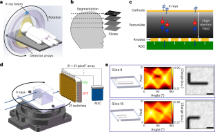
Indirect scintillator computed tomography (CT) imaging suffers from reduced image contrast and high-dose X-ray exposure due to inevitable light losses and multiple energy conversion steps. Here we report a direct lead-halide perovskite CT imager through low-cost spray-coating processes. Detector arrays with 980 μm absorber thickness and <10 nm surface roughness yield uniform X-ray response with detection quantum efficiency of 80% and noise-equivalent dose of 153 pGyair. The perovskite CT imager affords the three-dimensional reconstruction of a tooth under a low effective dose of 5.5 μSv, about two orders of magnitude smaller than dental cone-beam CT, and low-contrast detectability by resolving a 5 Hounsfield unit difference within a 5 mm region of interest.
中文翻译:
钙钛矿计算机断层扫描成像仪和三维重建
由于不可避免的光损失和多个能量转换步骤,间接闪烁体计算机断层扫描 (CT) 成像面临图像对比度降低和高剂量 X 射线暴露的问题。在这里,我们报告了一种通过低成本喷涂工艺直接卤化铅钙钛矿 CT 成像仪。吸收体厚度为 980 μm、表面粗糙度为 <10 nm 的探测器阵列可产生均匀的 X 射线响应,探测量子效率为 80%,噪声等效剂量为 153 pGy空气。钙钛矿 CT 成像仪可在 5.5 μSv 的低有效剂量下实现牙齿的三维重建,大约比牙科锥形束 CT 小两个数量级,并且通过在 5 秒内解析 5 亨斯菲尔德单位差异来实现低对比度可检测性。 mm 感兴趣的区域。


 京公网安备 11010802027423号