Our official English website, www.x-mol.net, welcomes your
feedback! (Note: you will need to create a separate account there.)
Fluorescent probes for neuroscience: imaging ex vivo brain tissue sections
Analyst ( IF 3.6 ) Pub Date : 2024-08-14 , DOI: 10.1039/d4an00663a Bradley J Schwehr 1 , David Hartnell 1, 2 , Gaewyn Ellison 1, 2 , Madison T Hindes 3 , Breah Milford 1 , Elena Dallerba 1 , Shane M Hickey 3 , Frederick M Pfeffer 4 , Doug A Brooks 3 , Massimiliano Massi 1 , Mark J Hackett 1, 2
Analyst ( IF 3.6 ) Pub Date : 2024-08-14 , DOI: 10.1039/d4an00663a Bradley J Schwehr 1 , David Hartnell 1, 2 , Gaewyn Ellison 1, 2 , Madison T Hindes 3 , Breah Milford 1 , Elena Dallerba 1 , Shane M Hickey 3 , Frederick M Pfeffer 4 , Doug A Brooks 3 , Massimiliano Massi 1 , Mark J Hackett 1, 2
Affiliation

|
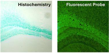
Neurobiological research relies heavily on imaging techniques, such as fluorescence microscopy, to understand neurological function and disease processes. However, the number and variety of fluorescent probes available for ex vivo tissue section imaging limits the advance of research in the field. In this review, we outline the current range of fluorescent probes that are available to researchers for ex vivo brain section imaging, including their physical and chemical characteristics, staining targets, and examples of discoveries for which they have been used. This review is organised into sections based on the biological target of the probe, including subcellular organelles, chemical species (e.g., labile metal ions), and pathological phenomenon (e.g., degenerating cells, aggregated proteins). We hope to inspire further development in this field, given the considerable benefits to be gained by the greater availability of suitably sensitive probes that have specificity for important brain tissue targets.
中文翻译:

用于神经科学的荧光探针:离体脑组织切片成像
神经生物学研究在很大程度上依赖于成像技术(例如荧光显微镜)来了解神经功能和疾病过程。然而,可用于离体组织切片成像的荧光探针的数量和种类限制了该领域研究的进展。在这篇综述中,我们概述了研究人员可用于离体脑切片成像的当前荧光探针范围,包括它们的物理和化学特性、染色目标以及它们已被使用的发现示例。本综述根据探针的生物学目标分为几个部分,包括亚细胞器、化学物质(例如不稳定的金属离子)和病理现象(例如退化细胞、聚集的蛋白质)。鉴于通过更多地提供对重要脑组织目标具有特异性的适当敏感的探针可以获得相当大的好处,我们希望激发该领域的进一步发展。
更新日期:2024-08-14
中文翻译:

用于神经科学的荧光探针:离体脑组织切片成像
神经生物学研究在很大程度上依赖于成像技术(例如荧光显微镜)来了解神经功能和疾病过程。然而,可用于离体组织切片成像的荧光探针的数量和种类限制了该领域研究的进展。在这篇综述中,我们概述了研究人员可用于离体脑切片成像的当前荧光探针范围,包括它们的物理和化学特性、染色目标以及它们已被使用的发现示例。本综述根据探针的生物学目标分为几个部分,包括亚细胞器、化学物质(例如不稳定的金属离子)和病理现象(例如退化细胞、聚集的蛋白质)。鉴于通过更多地提供对重要脑组织目标具有特异性的适当敏感的探针可以获得相当大的好处,我们希望激发该领域的进一步发展。


 京公网安备 11010802027423号