当前位置:
X-MOL 学术
›
Ann. N. Y. Acad. Sci.
›
论文详情
Our official English website, www.x-mol.net, welcomes your
feedback! (Note: you will need to create a separate account there.)
The causal role of the subthalamic nucleus in the inhibitory network
Annals of the New York Academy of Sciences ( IF 4.1 ) Pub Date : 2024-08-08 , DOI: 10.1111/nyas.15193 Ignacio Obeso 1 , Francis R Loayza 2, 3 , Rafael González-Redondo 4 , Federico Villagra 3 , Elkin Luis 3 , Marjan Jahanshahi 5 , José A Obeso 6, 7 , Maria A Pastor 3, 8
Annals of the New York Academy of Sciences ( IF 4.1 ) Pub Date : 2024-08-08 , DOI: 10.1111/nyas.15193 Ignacio Obeso 1 , Francis R Loayza 2, 3 , Rafael González-Redondo 4 , Federico Villagra 3 , Elkin Luis 3 , Marjan Jahanshahi 5 , José A Obeso 6, 7 , Maria A Pastor 3, 8
Affiliation
|
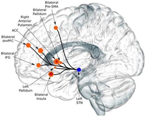
The neural network mediating successful response inhibition mainly includes right hemisphere activation of the pre-supplementary motor area, inferior frontal gyrus (IFG), subthalamic nucleus (STN), and caudate nucleus. However, the causal role of these regions in the inhibitory network is undefined. Five patients with Parkinson's disease were assessed prior to and after therapeutic thermal ablation of the right STN in two separate functional magnetic resonance imaging (fMRI) sessions while performing a stop-signal task. Initiation times were faster but motor inhibition with the left hand (contralateral to the lesion) was significantly impaired as evident in prolonged stop-signal reaction times. Reduced inhibition after right subthalamotomy was associated (during successful inhibition) with the recruitment of basal ganglia regions outside the established inhibitory network. They included the putamen and caudate together with the anterior cingulate cortex and IFG of the left hemisphere. Subsequent network connectivity analysis (with the seed over the nonlesioned left STN) revealed a new inhibitory network after right subthalamotomies. Our results highlight the causal role of the right STN in the neural network for motor inhibition and the possible basal ganglia mechanisms for compensation upon losing a key node of the inhibition network.
中文翻译:

丘脑底核在抑制网络中的因果作用
介导成功反应抑制的神经网络主要包括右半球前辅助运动区、额下回(IFG)、丘脑底核(STN)和尾状核的激活。然而,这些区域在抑制网络中的因果作用尚不清楚。在执行停止信号任务的同时,在两次独立的功能性磁共振成像 (fMRI) 过程中对 5 名帕金森病患者在右侧 STN 治疗性热消融之前和之后进行了评估。启动时间更快,但左手(病变对侧)的运动抑制明显受损,如停止信号反应时间延长所示。右侧丘脑底切开术后抑制的减少与已建立的抑制网络之外的基底神经节区域的募集有关(在成功抑制期间)。它们包括壳核和尾状核以及左半球的前扣带皮层和 IFG。随后的网络连接分析(种子位于未病变的左侧 STN 上)揭示了右侧丘脑底切除术后的新抑制网络。我们的结果强调了正确的 STN 在运动抑制神经网络中的因果作用,以及在失去抑制网络的关键节点时可能的基底神经节补偿机制。
更新日期:2024-08-08
中文翻译:
丘脑底核在抑制网络中的因果作用
介导成功反应抑制的神经网络主要包括右半球前辅助运动区、额下回(IFG)、丘脑底核(STN)和尾状核的激活。然而,这些区域在抑制网络中的因果作用尚不清楚。在执行停止信号任务的同时,在两次独立的功能性磁共振成像 (fMRI) 过程中对 5 名帕金森病患者在右侧 STN 治疗性热消融之前和之后进行了评估。启动时间更快,但左手(病变对侧)的运动抑制明显受损,如停止信号反应时间延长所示。右侧丘脑底切开术后抑制的减少与已建立的抑制网络之外的基底神经节区域的募集有关(在成功抑制期间)。它们包括壳核和尾状核以及左半球的前扣带皮层和 IFG。随后的网络连接分析(种子位于未病变的左侧 STN 上)揭示了右侧丘脑底切除术后的新抑制网络。我们的结果强调了正确的 STN 在运动抑制神经网络中的因果作用,以及在失去抑制网络的关键节点时可能的基底神经节补偿机制。


 京公网安备 11010802027423号