当前位置:
X-MOL 学术
›
ACS Appl. Mater. Interfaces
›
论文详情
Our official English website, www.x-mol.net, welcomes your
feedback! (Note: you will need to create a separate account there.)
Correction to “Wearable Magnetoelectric Stimulation for Chronic Wound Healing by Electrospun CoFe2O4@CTAB/PVDF Dressings”
ACS Applied Materials & Interfaces ( IF 8.3 ) Pub Date : 2024-07-23 , DOI: 10.1021/acsami.4c10024 Qi Ke , Xinyi Zhang , Yuan Yang , Qi Chen , Jianyu Su , Youhong Tang , Liming Fang
ACS Applied Materials & Interfaces ( IF 8.3 ) Pub Date : 2024-07-23 , DOI: 10.1021/acsami.4c10024 Qi Ke , Xinyi Zhang , Yuan Yang , Qi Chen , Jianyu Su , Youhong Tang , Liming Fang
|
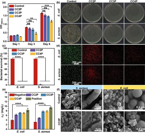
In the original version of the manuscript, Figure S9 (page 11 of the Supporting Information) contained partial repetition errors in certain cell viability images, Figure 3 (page 9842) and Figure S7 (page 9 of the Supporting Information) had spelling mistakes, and Figure 6 (page 9847) displayed some images with incorrect magnification scales. The figures and Supporting Information have been corrected. Figure 3. Antibacterial activity against E. coli and S. aureus and cytotoxicity analysis with different CTAB amounts. (a) CCK-8 results for cultured cells on fiber membranes with different CTAB contents. (b) Photos of colonies on plates. (c) Survival rates obtained from plate counts. (d) Fluorescent images of live (green)/dead (red) staining. (e) Potassium ion leakage in bacterial culture fluid after CTAB treatment. (f) SEM images of ruptured cell membranes. Note that the four symbols in the sample codes represent CoFe2O4, CTAB, the mass percentage of CTAB, and PVDF. Figure 6. Molecular mechanism of magnetoelectric stimulation dressings in promoting healing of infected wounds in diabetic rats. (a) Expression of factors related to wound healing on the seventh day postsurgery. (b) Quantitative analysis of blood vessels. (c) Quantitative analysis of VEGF, TGF-β, and IL, promoting vascular formation (CD31 marked in white triangles and VEGF marked in green) and extracellular matrix development (TGF-β marked in green), while suppressing the inflammatory phase (IL-6 marked in green). The Supporting Information is available free of charge at https://pubs.acs.org/doi/10.1021/acsami.4c10024. Schematic illustration of the CFO synthesis process and the improved crystal structure and enhanced magnetic property of CFO by annealing at higher temperatures (Figure S1), along with the lattice parameters and grain size of CFO (Table S1). Schematic illustration of the PVDF phase and molecular structures, β phase induced by electrospinning, and the polarization electric field curves of the PVDF fiber film, along with a schematic illustration of the piezoelectric nanogenerator working mechanism (Figure S2). Morphology and composition analysis of CCP (Figure S3). Simulation of the wearable magnetic field generator (Figure S4) and magnetoelectric coupling effect (Figure S5). Live/dead images of L929 cells cultured on CCP samples with different CTAB contents (Figure S6). Antibacterial effects of the magnetic field coupled with CCP film (Figure S7). Effects of fiber composition and magnetic field on the in vitro proliferation activity of L929 cells (Figure S8). Live/dead images of L929 cells cultured on samples with different compositions under various magnetic field stimulations (Figure S9). In vitro PCR under magnetic field stimulation (Figure S10). Primer sequences used in Q-RT-PCR (Table S2). Magnetic field stimulation environment of rat breeding and trace maps of the wound healing process (Figure S11) (PDF) Most electronic Supporting Information files are available without a subscription to ACS Web Editions. Such files may be downloaded by article for research use (if there is a public use license linked to the relevant article, that license may permit other uses). Permission may be obtained from ACS for other uses through requests via the RightsLink permission system: http://pubs.acs.org/page/copyright/permissions.html. This article has not yet been cited by other publications.
中文翻译:

对“静电纺丝 CoFe2O4@CTAB/PVDF 敷料用于慢性伤口愈合的可穿戴磁电刺激”的更正
在原稿的原始版本中,图S9(支持信息第11页)在某些细胞活力图像中包含部分重复错误,图3(第9842页)和图S7(支持信息第9页)有拼写错误,并且图 6(第 9847 页)显示了一些放大比例不正确的图像。数字和支持信息已更正。图 3. 不同 CTAB 量对大肠杆菌和金黄色葡萄球菌的抗菌活性和细胞毒性分析。 (a) 不同 CTAB 含量的纤维膜上培养细胞的 CCK-8 结果。 (b) 平板上菌落的照片。 (c) 从平板计数中获得的存活率。 (d) 活(绿色)/死(红色)染色的荧光图像。 (e) CTAB 处理后细菌培养液中钾离子渗漏。 (f) 破裂细胞膜的 SEM 图像。注意,示例代码中的四个符号分别代表CoFe 2 O 4 、CTAB、CTAB的质量百分比和PVDF。图6.磁电刺激敷料促进糖尿病大鼠感染伤口愈合的分子机制。 (a)术后第七天伤口愈合相关因子的表达。 (b) 血管的定量分析。 (c) VEGF、TGF-β 和 IL 的定量分析,促进血管形成(白色三角形标记的 CD31,绿色标记的 VEGF)和细胞外基质发育(绿色标记的 TGF-β),同时抑制炎症阶段(IL) -6 标记为绿色)。支持信息可在 https://pubs.acs.org/doi/10.1021/acsami.4c10024 免费获取。 CFO 合成过程和通过在较高温度下退火改善晶体结构和增强 CFO 磁性能的示意图(图 S1),以及 CFO 的晶格参数和晶粒尺寸(表 S1)。 PVDF相和分子结构、静电纺丝诱导的β相、PVDF纤维膜的极化电场曲线的示意图,以及压电纳米发电机工作机制的示意图(图S2)。 CCP的形态和成分分析(图S3)。可穿戴磁场发生器(图S4)和磁电耦合效应(图S5)的仿真。在具有不同 CTAB 含量的 CCP 样品上培养的 L929 细胞的活/死图像(图 S6)。磁场与 CCP 膜结合的抗菌效果(图 S7)。纤维成分和磁场对L929细胞体外增殖活性的影响(图S8)。在不同磁场刺激下,在不同成分的样品上培养的 L929 细胞的活/死图像(图 S9)。磁场刺激下的体外 PCR(图 S10)。 Q-RT-PCR 中使用的引物序列(表 S2)。大鼠繁殖的磁场刺激环境和伤口愈合过程的轨迹图(图 S11)(PDF) 大多数电子支持信息文件无需订阅 ACS 网络版即可获得。此类文件可以按文章下载用于研究用途(如果有链接到相关文章的公共使用许可证,则该许可证可能允许其他用途)。可以通过 RightsLink 许可系统提出请求,从 ACS 获得许可用于其他用途:http://pubs.acs.org/page/copyright/permissions.html。 这篇文章尚未被其他出版物引用。
更新日期:2024-07-23
中文翻译:
对“静电纺丝 CoFe2O4@CTAB/PVDF 敷料用于慢性伤口愈合的可穿戴磁电刺激”的更正
在原稿的原始版本中,图S9(支持信息第11页)在某些细胞活力图像中包含部分重复错误,图3(第9842页)和图S7(支持信息第9页)有拼写错误,并且图 6(第 9847 页)显示了一些放大比例不正确的图像。数字和支持信息已更正。图 3. 不同 CTAB 量对大肠杆菌和金黄色葡萄球菌的抗菌活性和细胞毒性分析。 (a) 不同 CTAB 含量的纤维膜上培养细胞的 CCK-8 结果。 (b) 平板上菌落的照片。 (c) 从平板计数中获得的存活率。 (d) 活(绿色)/死(红色)染色的荧光图像。 (e) CTAB 处理后细菌培养液中钾离子渗漏。 (f) 破裂细胞膜的 SEM 图像。注意,示例代码中的四个符号分别代表CoFe 2 O 4 、CTAB、CTAB的质量百分比和PVDF。图6.磁电刺激敷料促进糖尿病大鼠感染伤口愈合的分子机制。 (a)术后第七天伤口愈合相关因子的表达。 (b) 血管的定量分析。 (c) VEGF、TGF-β 和 IL 的定量分析,促进血管形成(白色三角形标记的 CD31,绿色标记的 VEGF)和细胞外基质发育(绿色标记的 TGF-β),同时抑制炎症阶段(IL) -6 标记为绿色)。支持信息可在 https://pubs.acs.org/doi/10.1021/acsami.4c10024 免费获取。 CFO 合成过程和通过在较高温度下退火改善晶体结构和增强 CFO 磁性能的示意图(图 S1),以及 CFO 的晶格参数和晶粒尺寸(表 S1)。 PVDF相和分子结构、静电纺丝诱导的β相、PVDF纤维膜的极化电场曲线的示意图,以及压电纳米发电机工作机制的示意图(图S2)。 CCP的形态和成分分析(图S3)。可穿戴磁场发生器(图S4)和磁电耦合效应(图S5)的仿真。在具有不同 CTAB 含量的 CCP 样品上培养的 L929 细胞的活/死图像(图 S6)。磁场与 CCP 膜结合的抗菌效果(图 S7)。纤维成分和磁场对L929细胞体外增殖活性的影响(图S8)。在不同磁场刺激下,在不同成分的样品上培养的 L929 细胞的活/死图像(图 S9)。磁场刺激下的体外 PCR(图 S10)。 Q-RT-PCR 中使用的引物序列(表 S2)。大鼠繁殖的磁场刺激环境和伤口愈合过程的轨迹图(图 S11)(PDF) 大多数电子支持信息文件无需订阅 ACS 网络版即可获得。此类文件可以按文章下载用于研究用途(如果有链接到相关文章的公共使用许可证,则该许可证可能允许其他用途)。可以通过 RightsLink 许可系统提出请求,从 ACS 获得许可用于其他用途:http://pubs.acs.org/page/copyright/permissions.html。 这篇文章尚未被其他出版物引用。


 京公网安备 11010802027423号