Nature Genetics ( IF 31.7 ) Pub Date : 2024-07-24 , DOI: 10.1038/s41588-024-01802-x Amin Abedini , Jonathan Levinsohn , Konstantin A. Klötzer , Bernhard Dumoulin , Ziyuan Ma , Julia Frederick , Poonam Dhillon , Michael S. Balzer , Rojesh Shrestha , Hongbo Liu , Steven Vitale , Andi M. Bergeson , Kishor Devalaraja-Narashimha , Paola Grandi , Tanmoy Bhattacharyya , Erding Hu , Steven S. Pullen , Carine M. Boustany-Kari , Paolo Guarnieri , Anil Karihaloo , Daniel Traum , Hanying Yan , Kyle Coleman , Matthew Palmer , Lea Sarov-Blat , Lori Morton , Christopher A. Hunter , Klaus H. Kaestner , Mingyao Li , Katalin Susztak
|
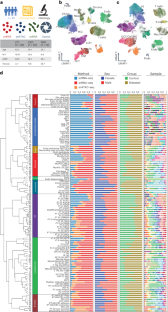
Kidneys are intricate three-dimensional structures in the body, yet the spatial and molecular principles of kidney health and disease remain inadequately understood. We generated high-quality datasets for 81 samples, including single-cell, single-nuclear, spot-level (Visium) and single-cell resolution (CosMx) spatial-RNA expression and single-nuclear open chromatin, capturing cells from healthy, diabetic and hypertensive diseased human kidneys. Combining these data, we identify cell types and map them to their locations within the tissue. Unbiased deconvolution of the spatial data identifies the following four distinct microenvironments: glomerular, immune, tubule and fibrotic. We describe the complex organization of microenvironments in health and disease and find that the fibrotic microenvironment is able to molecularly classify human kidneys and offers an improved prognosis compared to traditional histopathology. We provide a comprehensive spatially resolved molecular roadmap of the human kidney and the fibrotic process, demonstrating the clinical utility of spatial transcriptomics.
中文翻译:
人类肾脏的单细胞多组学和空间分析表明肾脏疾病进展中的纤维化微环境
肾脏是体内复杂的三维结构,但肾脏健康和疾病的空间和分子原理仍然没有得到充分的了解。我们为 81 个样本生成了高质量数据集,包括单细胞、单核、点级 (Visium) 和单细胞分辨率 (CosMx) 空间 RNA 表达和单核开放染色质,捕获来自健康、糖尿病患者的细胞和高血压患病的人类肾脏。结合这些数据,我们识别细胞类型并将它们映射到组织内的位置。空间数据的无偏反卷积识别出以下四种不同的微环境:肾小球、免疫、肾小管和纤维化。我们描述了健康和疾病中微环境的复杂组织,发现纤维化微环境能够对人类肾脏进行分子分类,并与传统的组织病理学相比提供更好的预后。我们提供了人类肾脏和纤维化过程的全面空间解析分子路线图,展示了空间转录组学的临床实用性。


 京公网安备 11010802027423号