Our official English website, www.x-mol.net, welcomes your
feedback! (Note: you will need to create a separate account there.)
Rigidity percolation and active advection synergize in the actomyosin cortex to drive amoeboid cell motility
Developmental Cell ( IF 10.7 ) Pub Date : 2024-07-23 , DOI: 10.1016/j.devcel.2024.06.023 Juan Manuel García-Arcos 1 , Johannes Ziegler 2 , Silvia Grigolon 3 , Loïc Reymond 4 , Gaurav Shajepal 5 , Cédric J Cattin 6 , Alexis Lomakin 7 , Daniel J Müller 6 , Verena Ruprecht 8 , Stefan Wieser 2 , Raphael Voituriez 9 , Matthieu Piel 1
Developmental Cell ( IF 10.7 ) Pub Date : 2024-07-23 , DOI: 10.1016/j.devcel.2024.06.023 Juan Manuel García-Arcos 1 , Johannes Ziegler 2 , Silvia Grigolon 3 , Loïc Reymond 4 , Gaurav Shajepal 5 , Cédric J Cattin 6 , Alexis Lomakin 7 , Daniel J Müller 6 , Verena Ruprecht 8 , Stefan Wieser 2 , Raphael Voituriez 9 , Matthieu Piel 1
Affiliation

|
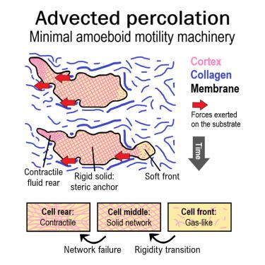
Spontaneous locomotion is a common feature of most metazoan cells, generally attributed to the properties of actomyosin networks. This force-producing machinery has been studied down to the most minute molecular details, especially in lamellipodium-driven migration. Nevertheless, how actomyosin networks work inside contraction-driven amoeboid cells still lacks unifying principles. Here, using stable motile blebs from HeLa cells as a model amoeboid motile system, we imaged the dynamics of the actin cortex at the single filament level and revealed the co-existence of three distinct rheological phases. We introduce “advected percolation,” a process where rigidity percolation and active advection synergize, spatially organizing the actin network’s mechanical properties into a minimal and generic locomotion mechanism. Expanding from our observations on simplified systems, we speculate that this model could explain, down to the single actin filament level, how amoeboid cells, such as cancer or immune cells, can propel efficiently through complex 3D environments.
中文翻译:

肌动球蛋白皮层中的刚性渗流和主动平流协同作用,以驱动变形虫细胞运动
自发运动是大多数后生动物细胞的共同特征,通常归因于肌动球蛋白网络的特性。这种产生力的机制已经被研究到最微小的分子细节,特别是在片状足驱动的迁移中。然而,肌动球蛋白网络如何在收缩驱动的变形虫细胞内工作仍然缺乏统一的原则。在这里,使用来自 HeLa 细胞的稳定运动气泡作为模型变形虫运动系统,我们在单丝水平上对肌动蛋白皮层的动力学进行了成像,并揭示了三个不同流变阶段的共存。我们引入了“平流渗流”,这是一个刚性渗流和主动平流协同作用的过程,在空间上将肌动蛋白网络的机械特性组织成最小和通用的运动机制。从我们对简化系统的观察中扩展,我们推测该模型可以解释,直到单个肌动蛋白丝水平,变形虫细胞(如癌症或免疫细胞)如何在复杂的 3D 环境中有效推动。
更新日期:2024-07-23
中文翻译:

肌动球蛋白皮层中的刚性渗流和主动平流协同作用,以驱动变形虫细胞运动
自发运动是大多数后生动物细胞的共同特征,通常归因于肌动球蛋白网络的特性。这种产生力的机制已经被研究到最微小的分子细节,特别是在片状足驱动的迁移中。然而,肌动球蛋白网络如何在收缩驱动的变形虫细胞内工作仍然缺乏统一的原则。在这里,使用来自 HeLa 细胞的稳定运动气泡作为模型变形虫运动系统,我们在单丝水平上对肌动蛋白皮层的动力学进行了成像,并揭示了三个不同流变阶段的共存。我们引入了“平流渗流”,这是一个刚性渗流和主动平流协同作用的过程,在空间上将肌动蛋白网络的机械特性组织成最小和通用的运动机制。从我们对简化系统的观察中扩展,我们推测该模型可以解释,直到单个肌动蛋白丝水平,变形虫细胞(如癌症或免疫细胞)如何在复杂的 3D 环境中有效推动。


 京公网安备 11010802027423号