当前位置:
X-MOL 学术
›
ACS Appl. Mater. Interfaces
›
论文详情
Our official English website, www.x-mol.net, welcomes your
feedback! (Note: you will need to create a separate account there.)
Driving Osteocytogenesis from Mesenchymal Stem Cells in Osteon-like Biomimetic Nanofibrous Scaffolds
ACS Applied Materials & Interfaces ( IF 8.3 ) Pub Date : 2024-07-23 , DOI: 10.1021/acsami.3c14785 Farhad Soheilmoghaddam 1, 2 , Hadi Hezaveh 1 , Madeleine Rumble 2 , Justin J Cooper-White 1, 2
ACS Applied Materials & Interfaces ( IF 8.3 ) Pub Date : 2024-07-23 , DOI: 10.1021/acsami.3c14785 Farhad Soheilmoghaddam 1, 2 , Hadi Hezaveh 1 , Madeleine Rumble 2 , Justin J Cooper-White 1, 2
Affiliation
|
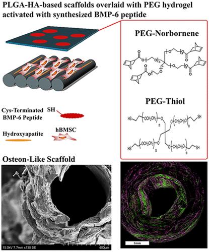
The treatment of critical-sized bone defects caused by tumor removal, skeletal injuries, or infections continues to pose a major clinical challenge. A popular potential alternative solution to autologous bone grafts is a tissue-engineered approach that utilizes the combination of mesenchymal stromal/stem cells (MSCs) with synthetic biomaterial scaffolds. This approach aims to support new bone formation by mimicking many of the biochemical and biophysical cues present within native bone. Regrettably, osteocyte cells, crucial for bone maturation and homeostasis, are rarely produced within MSC-seeded scaffolds, thereby restricting the development of fully mature cortical bone from these synthetic implants. In this work, we have constructed a multimodal scaffold by combining electrospun poly(lactic-co-glycolic acid) (PLGA) fibrous scaffolds with poly(ethylene glycol) (PEG)-based hydrogels that mimic the functional unit of cortical bone, osteon (osteon-mimetic) scaffolds. These scaffolds were decorated with a novel bone morphogenic protein-6 (BMP6) peptide (BMP6p) after our findings revealed that the BMP6p drives higher levels of Smad signaling than the full-length protein counterpart, soluble or when bound to the PEG hydrogel backbone. We show that our osteon-mimetic scaffolds, in presenting concentric layers of BMP6p-PEG hydrogel overlaid on MSC-seeded PLGA nanofibers, promoted the rapid formation of osteocyte-like cells with a phenotypic dendritic morphology, producing early osteocyte markers, including E11/gp38 (E11). Maturation of these osteocyte-like cells was further confirmed by the observation of significant dentin matrix protein 1 (DMP1) throughout our bilayered scaffolds after 3 weeks, even when cultured in a medium without dexamethasone (DEX) or any other osteogenic supplements. These results demonstrate that these osteon-mimetic scaffolds, in presenting biochemical and topographical cues reminiscent of the forming osteon, can drive the formation of osteocyte-like cells in vitro from hBMSCs without the need for any osteogenic factor media supplementation.
中文翻译:

在类骨仿生纳米纤维支架中驱动间充质干细胞的骨细胞生成
由肿瘤切除、骨骼损伤或感染引起的临界骨缺损的治疗仍然是一项重大的临床挑战。自体骨移植的一种流行的潜在替代解决方案是组织工程方法,该方法利用间充质基质/干细胞(MSC)与合成生物材料支架的组合。这种方法旨在通过模仿天然骨中存在的许多生化和生物物理线索来支持新骨的形成。遗憾的是,对于骨成熟和体内平衡至关重要的骨细胞很少在 MSC 接种的支架内产生,从而限制了这些合成植入物发育完全成熟的皮质骨。在这项工作中,我们通过将电纺聚乳酸乙醇酸(PLGA) 纤维支架与基于聚乙二醇 (PEG) 的水凝胶相结合,构建了一种多模态支架,该水凝胶模仿了皮质骨的功能单元,骨。仿骨)支架。我们的研究结果表明,与全长蛋白质对应物(可溶或与 PEG 水凝胶骨架结合时)相比,BMP6p 能驱动更高水平的 Smad 信号传导,这些支架上装饰有新型骨形态发生蛋白 6 (BMP6) 肽 (BMP6p)。我们表明,我们的骨模拟支架呈现出覆盖在 MSC 接种的 PLGA 纳米纤维上的 BMP6p-PEG 水凝胶同心层,促进了具有表型树突形态的骨细胞样细胞的快速形成,产生早期骨细胞标记物,包括 E11/gp38 (E11)。 3周后,即使在不含地塞米松 (DEX) 或任何其他成骨补充剂的培养基中培养,通过在我们的双层支架中观察到显着的牙本质基质蛋白 1 (DMP1),进一步证实了这些骨细胞样细胞的成熟。这些结果表明,这些仿骨支架呈现出让人想起形成骨的生化和地形线索,可以在体外驱动 hBMSC 形成骨细胞样细胞,而不需要补充任何成骨因子培养基。
更新日期:2024-07-23
中文翻译:
在类骨仿生纳米纤维支架中驱动间充质干细胞的骨细胞生成
由肿瘤切除、骨骼损伤或感染引起的临界骨缺损的治疗仍然是一项重大的临床挑战。自体骨移植的一种流行的潜在替代解决方案是组织工程方法,该方法利用间充质基质/干细胞(MSC)与合成生物材料支架的组合。这种方法旨在通过模仿天然骨中存在的许多生化和生物物理线索来支持新骨的形成。遗憾的是,对于骨成熟和体内平衡至关重要的骨细胞很少在 MSC 接种的支架内产生,从而限制了这些合成植入物发育完全成熟的皮质骨。在这项工作中,我们通过将电纺聚乳酸乙醇酸(PLGA) 纤维支架与基于聚乙二醇 (PEG) 的水凝胶相结合,构建了一种多模态支架,该水凝胶模仿了皮质骨的功能单元,骨。仿骨)支架。我们的研究结果表明,与全长蛋白质对应物(可溶或与 PEG 水凝胶骨架结合时)相比,BMP6p 能驱动更高水平的 Smad 信号传导,这些支架上装饰有新型骨形态发生蛋白 6 (BMP6) 肽 (BMP6p)。我们表明,我们的骨模拟支架呈现出覆盖在 MSC 接种的 PLGA 纳米纤维上的 BMP6p-PEG 水凝胶同心层,促进了具有表型树突形态的骨细胞样细胞的快速形成,产生早期骨细胞标记物,包括 E11/gp38 (E11)。 3周后,即使在不含地塞米松 (DEX) 或任何其他成骨补充剂的培养基中培养,通过在我们的双层支架中观察到显着的牙本质基质蛋白 1 (DMP1),进一步证实了这些骨细胞样细胞的成熟。这些结果表明,这些仿骨支架呈现出让人想起形成骨的生化和地形线索,可以在体外驱动 hBMSC 形成骨细胞样细胞,而不需要补充任何成骨因子培养基。


 京公网安备 11010802027423号