当前位置:
X-MOL 学术
›
J. Med. Chem.
›
论文详情
Our official English website, www.x-mol.net, welcomes your feedback! (Note: you will need to create a separate account there.)
Correction to “Discovery of Novel Acridane-Based Tubulin Polymerization Inhibitors with Anticancer and Potential Immunomodulatory Effects”
Journal of Medicinal Chemistry ( IF 6.8 ) Pub Date : 2024-07-18 , DOI: 10.1021/acs.jmedchem.4c01498 Xiaopeng Peng , Yichang Ren , Wanyi Pan , Jin Liu , Jianjun Chen
Journal of Medicinal Chemistry ( IF 6.8 ) Pub Date : 2024-07-18 , DOI: 10.1021/acs.jmedchem.4c01498 Xiaopeng Peng , Yichang Ren , Wanyi Pan , Jin Liu , Jianjun Chen
|
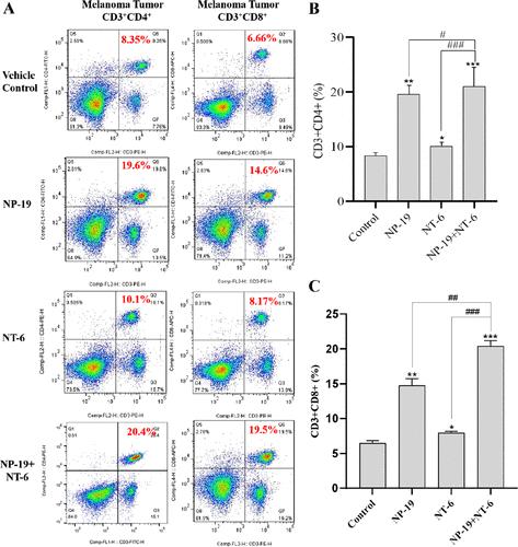
Page 634. During the assembly of Figure 12 in the original manuscript, due to the multitude of experimental groups and the relatively small size of the flow cytometry images, we inadvertently used a wrong image for the CD3+CD4+ T cell infiltration (rate = 26.3%) in the NP-19 + NT-6 combination therapy group (bottom left of panel A). Upon recent and careful review of the original manuscript, we identified this error and herein replace the wrong image with the correct one which shows a CD3+CD4+ T cell infiltration rate of 20.4% for the NP-19 + NT-6 combination therapy group. This corrected result still demonstrates that NT-6 can enhance T cell infiltration in the presence of the PD-L1 small molecule inhibitor NP-19, and does not change the scientific conclusions of the article in any way. Page 635. The text associated with Figure 12 in the first paragraph of section 2.2.11 should be modified as follows: To further explore the immunomodulatory mechanism of NT-6, flow cytometry was utilized to measure the tumor-infiltrating lymphocytes (TILs) in melanoma tumor tissues. As shown in Figure 12, the percentages of CD3+CD4+ cells (activated helper T cells) in the melanoma tumor tissues of the three treatment groups (NP-19, NT-6, NP-19 + NT-6) were 19.6, 10.1, and 20.4%, respectively, as compared to the vehicle control group (8.35%). Additionally, the percentages of CD3+CD8+ cells (activated cytotoxic T cells) in the melanoma tumor tissues of the three treatment groups (NP-19, NT-6, NP-19 + NT-6) were 14.6, 8.17, and 19.5%, respectively, as compared to 6.66% for the vehicle control group. Although the percentages of CD3+CD4+ cells (10.1%) and CD3+CD8+ cells (8.17%) in the NT-6 treated group were only slightly higher than the vehicle control group (8.35, 6.66% for CD3+CD4+ cells and CD3+CD8+ cells, respectively), the percentages of CD3+CD4+ cells (20.4%) and CD3+CD8+ cells (19.5%) were increased in the combination group (NP-19 + NT-6). Figure 12. Detection of infiltrating lymphocytes in melanoma tumor tissues by flow cytometry. (A) Representative images for CD3+CD4+ cells (helper T cells) and CD3+CD8+ cells (activated cytotoxic T cells) in tumor tissues of vehicle control, NP-19, NT-6, and (NP-19 + NT-6) combination-treated mice; melanoma tumor CD3+CD4+ (B) and CD3+CD8+ (C) cells in vehicle control, NP-19, NT-6, and (NP-19 + NT-6) combination-treated mice. ***P < 0.001, **P < 0.01, *P < 0.05 compared with vehicle group (n = 6, Dunnett’s multiple comparison test), ###P < 0.001, #P < 0.05 compared with (NP-19 + NT-6)-treated group. All authors have agreed to the changes. We apologize for any confusion this may have caused and appreciate readers’ understanding as we make these necessary corrections to ensure the integrity of our research findings. This article has not yet been cited by other publications.
中文翻译:

对“具有抗癌和潜在免疫调节作用的新型吖啶基微管蛋白聚合抑制剂的发现”的更正
第634页。在原稿图12的组装过程中,由于实验组众多且流式细胞术图像尺寸相对较小,我们无意中对CD3 + CD4 <使用了错误的图像b1> NP-19 + NT-6 联合治疗组中的 T 细胞浸润(比率 = 26.3%)(A 组左下)。经过最近仔细审查原稿,我们发现了这个错误,并在此用正确的图像替换了错误的图像,显示 CD3 + CD4 + T 细胞浸润率为 20.4%为NP-19+NT-6联合治疗组。这个修正后的结果仍然证明NT-6在PD-L1小分子抑制剂NP-19存在的情况下可以增强T细胞浸润,并且不会以任何方式改变文章的科学结论。第635页。第2.2.11节第一段中与图12相关的文字应修改如下:为进一步探讨NT-6的免疫调节机制,采用流式细胞术测量肿瘤浸润淋巴细胞(TIL)黑色素瘤肿瘤组织。如图12所示,三个治疗组(NP-19、NT-6)黑色素瘤肿瘤组织中CD3 + CD4 + 细胞(活化的辅助T细胞)的百分比、NP-19 + NT-6)与载体对照组(8.35%)相比分别为 19.6%、10.1% 和 20.4%。此外,三个治疗组(NP-19、NT-6、NP-19)黑色素瘤肿瘤组织中 CD3 + CD8 + 细胞(活化的细胞毒性 T 细胞)的百分比+ NT-6)分别为 14.6、8.17 和 19.5%,而载体对照组为 6.66%。尽管 CD3 + CD4 + 细胞的百分比 (10.1%) 和 CD3 + CD8 + 细胞的百分比 (8.NT-6 治疗组中的 17%)仅略高于载体对照组(CD3 + CD4 + 细胞和 CD3 + CD8 + 细胞,分别)、CD3 + CD4 + 细胞的百分比 (20.4%) 和 CD3 + CD8 + 联合组(NP-19 + NT-6)细胞(19.5%)增加。图12.通过流式细胞术检测黑色素瘤肿瘤组织中的浸润淋巴细胞。 (A) CD3 + CD4 + 细胞(辅助性 T 细胞)和 CD3 + CD8 + 细胞(激活的细胞毒性 T 细胞)的代表性图像细胞)在载体对照、NP-19、NT-6和(NP-19 + NT-6)联合治疗的小鼠的肿瘤组织中;黑色素瘤肿瘤 CD3 + CD4 + (B) 和 CD3 + CD8 + (C) 载体对照细胞,NP-19, NT-6 和 (NP-19 + NT-6) 联合治疗的小鼠。 ***P < 0.001,**P < 0.01,*P < 0.05 与赋形剂组相比(n = 6,Dunnett 多重比较检验), ### P < 0.001, # 与(NP-19 + NT-6)治疗组相比,P < 0.05。所有作者均同意这些更改。对于这可能造成的任何混乱,我们深表歉意,并感谢读者的理解,因为我们会进行必要的更正,以确保我们研究结果的完整性。这篇文章尚未被其他出版物引用。
更新日期:2024-07-23
中文翻译:
对“具有抗癌和潜在免疫调节作用的新型吖啶基微管蛋白聚合抑制剂的发现”的更正
第634页。在原稿图12的组装过程中,由于实验组众多且流式细胞术图像尺寸相对较小,我们无意中对CD3 + CD4 <使用了错误的图像b1> NP-19 + NT-6 联合治疗组中的 T 细胞浸润(比率 = 26.3%)(A 组左下)。经过最近仔细审查原稿,我们发现了这个错误,并在此用正确的图像替换了错误的图像,显示 CD3 + CD4 + T 细胞浸润率为 20.4%为NP-19+NT-6联合治疗组。这个修正后的结果仍然证明NT-6在PD-L1小分子抑制剂NP-19存在的情况下可以增强T细胞浸润,并且不会以任何方式改变文章的科学结论。第635页。第2.2.11节第一段中与图12相关的文字应修改如下:为进一步探讨NT-6的免疫调节机制,采用流式细胞术测量肿瘤浸润淋巴细胞(TIL)黑色素瘤肿瘤组织。如图12所示,三个治疗组(NP-19、NT-6)黑色素瘤肿瘤组织中CD3 + CD4 + 细胞(活化的辅助T细胞)的百分比、NP-19 + NT-6)与载体对照组(8.35%)相比分别为 19.6%、10.1% 和 20.4%。此外,三个治疗组(NP-19、NT-6、NP-19)黑色素瘤肿瘤组织中 CD3 + CD8 + 细胞(活化的细胞毒性 T 细胞)的百分比+ NT-6)分别为 14.6、8.17 和 19.5%,而载体对照组为 6.66%。尽管 CD3 + CD4 + 细胞的百分比 (10.1%) 和 CD3 + CD8 + 细胞的百分比 (8.NT-6 治疗组中的 17%)仅略高于载体对照组(CD3 + CD4 + 细胞和 CD3 + CD8 + 细胞,分别)、CD3 + CD4 + 细胞的百分比 (20.4%) 和 CD3 + CD8 + 联合组(NP-19 + NT-6)细胞(19.5%)增加。图12.通过流式细胞术检测黑色素瘤肿瘤组织中的浸润淋巴细胞。 (A) CD3 + CD4 + 细胞(辅助性 T 细胞)和 CD3 + CD8 + 细胞(激活的细胞毒性 T 细胞)的代表性图像细胞)在载体对照、NP-19、NT-6和(NP-19 + NT-6)联合治疗的小鼠的肿瘤组织中;黑色素瘤肿瘤 CD3 + CD4 + (B) 和 CD3 + CD8 + (C) 载体对照细胞,NP-19, NT-6 和 (NP-19 + NT-6) 联合治疗的小鼠。 ***P < 0.001,**P < 0.01,*P < 0.05 与赋形剂组相比(n = 6,Dunnett 多重比较检验), ### P < 0.001, # 与(NP-19 + NT-6)治疗组相比,P < 0.05。所有作者均同意这些更改。对于这可能造成的任何混乱,我们深表歉意,并感谢读者的理解,因为我们会进行必要的更正,以确保我们研究结果的完整性。这篇文章尚未被其他出版物引用。


 京公网安备 11010802027423号