Nature Communications ( IF 14.7 ) Pub Date : 2024-07-20 , DOI: 10.1038/s41467-024-50285-1 Xinyi Zhang 1, 2 , Saradha Venkatachalapathy 3, 4 , Daniel Paysan 3, 4 , Paulina Schaerer 3, 4 , Claudio Tripodo 5, 6 , Caroline Uhler 1, 2 , G V Shivashankar 3, 4
|
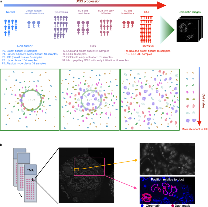
Ductal carcinoma in situ (DCIS) is a pre-invasive tumor that can progress to invasive breast cancer, a leading cause of cancer death. We generate a large-scale tissue microarray dataset of chromatin images, from 560 samples from 122 female patients in 3 disease stages and 11 phenotypic categories. Using representation learning on chromatin images alone, without multiplexed staining or high-throughput sequencing, we identify eight morphological cell states and tissue features marking DCIS. All cell states are observed in all disease stages with different proportions, indicating that cell states enriched in invasive cancer exist in small fractions in normal breast tissue. Tissue-level analysis reveals significant changes in the spatial organization of cell states across disease stages, which is predictive of disease stage and phenotypic category. Taken together, we show that chromatin imaging represents a powerful measure of cell state and disease stage of DCIS, providing a simple and effective tumor biomarker.
中文翻译:
染色质图像的无监督表示学习可识别 DCIS 中细胞状态和组织组织的变化
导管原位癌 (DCIS) 是一种浸润前肿瘤,可进展为浸润性乳腺癌,这是癌症死亡的主要原因。我们从来自 3 个疾病阶段和 11 个表型类别的 122 名女性患者的 560 个样本中生成了大规模染色质图像组织微阵列数据集。仅使用染色质图像上的表示学习,无需多重染色或高通量测序,我们就识别出标记 DCIS 的八种形态细胞状态和组织特征。在所有疾病阶段都观察到不同比例的所有细胞状态,表明在正常乳腺组织中存在少量浸润性癌症中富含的细胞状态。组织水平分析揭示了疾病阶段细胞状态空间组织的显着变化,这可以预测疾病阶段和表型类别。综上所述,我们发现染色质成像是衡量导管原位癌细胞状态和疾病阶段的有力手段,提供了一种简单而有效的肿瘤生物标志物。


 京公网安备 11010802027423号