Nature Communications ( IF 14.7 ) Pub Date : 2024-07-19 , DOI: 10.1038/s41467-024-50438-2 Lies van Baarle , Veronica De Simone , Linda Schneider , Sneha Santhosh , Saeed Abdurahiman , Francesca Biscu , Reiner Schneider , Lisa Zanoletti , Renata Siqueira de Mello , Sara Verbandt , Zedong Hu , Michelle Stakenborg , Bo-Jun Ke , Nathalie Stakenborg , Raquel Salvador Laureano , Balbina García-Reyes , Jonas Henn , Marieta Toma , Maxime Vanmechelen , Guy Boeckxstaens , Frederik De Smet , Abhishek D. Garg , Sales Ibiza , Sabine Tejpar , Sven Wehner , Gianluca Matteoli
|
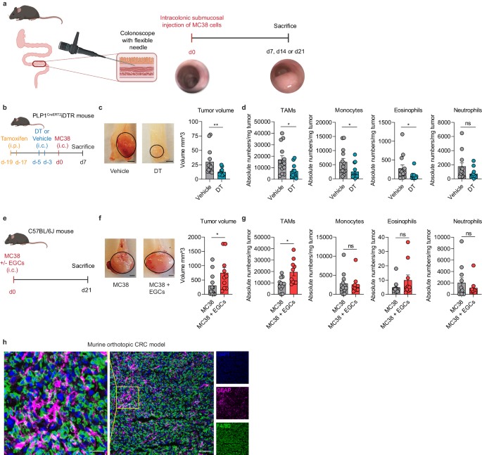
Enteric glia have been recently recognized as key components of the colonic tumor microenvironment indicating their potential role in colorectal cancer pathogenesis. Although enteric glia modulate immune responses in other intestinal diseases, their interaction with the colorectal cancer immune cell compartment remains unclear. Through a combination of single-cell and bulk RNA-sequencing, both in murine models and patients, here we find that enteric glia acquire an immunomodulatory phenotype by bi-directional communication with tumor-infiltrating monocytes. The latter direct a reactive enteric glial cell phenotypic and functional switch via glial IL-1R signaling. In turn, tumor glia promote monocyte differentiation towards pro-tumorigenic SPP1+ tumor-associated macrophages by IL-6 release. Enteric glia cell abundancy correlates with worse disease outcomes in preclinical models and colorectal cancer patients. Thereby, our study reveals a neuroimmune interaction between enteric glia and tumor-associated macrophages in the colorectal tumor microenvironment, providing insights into colorectal cancer pathogenesis.
中文翻译:
IL-1R信号驱动结直肠癌中肠胶质细胞-巨噬细胞的相互作用
肠神经胶质细胞最近被认为是结肠肿瘤微环境的关键组成部分,表明它们在结直肠癌发病机制中的潜在作用。尽管肠神经胶质细胞调节其他肠道疾病的免疫反应,但它们与结直肠癌免疫细胞区室的相互作用仍不清楚。通过结合单细胞和批量 RNA 测序,在小鼠模型和患者中,我们发现肠胶质细胞通过与肿瘤浸润单核细胞的双向通讯获得免疫调节表型。后者通过神经胶质细胞 IL-1R 信号传导指导反应性肠神经胶质细胞表型和功能转换。反过来,肿瘤胶质细胞通过 IL-6 释放促进单核细胞分化为促肿瘤 SPP1 + 肿瘤相关巨噬细胞。肠神经胶质细胞丰度与临床前模型和结直肠癌患者的较差疾病结果相关。因此,我们的研究揭示了结直肠肿瘤微环境中肠胶质细胞和肿瘤相关巨噬细胞之间的神经免疫相互作用,为结直肠癌发病机制提供了见解。


 京公网安备 11010802027423号