Our official English website, www.x-mol.net, welcomes your feedback! (Note: you will need to create a separate account there.)
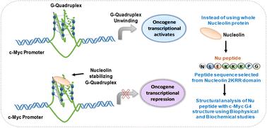
Structural analysis of peptide identified from the 2KRR domain of the nucleolin protein with a c-Myc G4 structure using biophysical and biochemical methods
RSC Advances ( IF 3.9 ) Pub Date : 2024-07-19 , DOI: 10.1039/d4ra02785j Sarvpreet Kaur 1 , Nikita Kundu 1 , Taniya Sharma 1 , J. Shankaraswamy 2 , Sweta Singh 3 , Sarika Saxena 1
RSC Advances ( IF 3.9 ) Pub Date : 2024-07-19 , DOI: 10.1039/d4ra02785j Sarvpreet Kaur 1 , Nikita Kundu 1 , Taniya Sharma 1 , J. Shankaraswamy 2 , Sweta Singh 3 , Sarika Saxena 1
Affiliation
|
For the first time, the c-Myc G4 structure is reported to be stabilized by binding of the peptide (derived from the 2KRR domain of the nucleolin protein called the Nu peptide) in the loop region of the G-quadruplex structure by stacking interactions. CD results showed the formation of parallel G4 structure in the presence of 100 mM Na+ or 100 mM K+ with the appearance of two isodichroic points at 229 nm, 254 nm and 252 nm in the presence of 100 mM Na+ or 100 mM K+, respectively. In addition, in UV thermal and CD melting studies, we observed drastic changes with an increase in the hyperchromicity at a DNA : peptide ratio of 1 : 50. On titrating the Nu peptide with c-Myc G4, we calculated the value of binding constant (Ka) by plotting fluorescence intensity and DNA concentration as 0.1369 ± 0.008 μM and 0.1277 ± 0.073 μM in Na+ and K+, respectively, which confirms the strong association of Nu peptide with c-Myc G4. The Nu peptide showed preferential cytotoxicity against MDA-MB-231 cells with IC50 values of 5.020 μM and 5.501 μM after 72 and 96 hours. This approach suggests a novel strategy to target G4 structure using natural key peptide segments derived from G4 stabilizing protein.
中文翻译:

使用生物物理和生化方法对从具有 c-Myc G4 结构的核仁蛋白的 2KRR 结构域中鉴定出的肽进行结构分析
首次报道,c-Myc G4 结构通过肽(源自称为 Nu 肽的核仁蛋白的 2KRR 结构域)通过堆积相互作用结合在 G-四链体结构的环区域中而稳定。 CD结果表明,在100 mM Na + 或100 mM K + 存在下形成平行的G4结构,并在229 nm、254 nm和252 nm处出现两个等色点分别在 100 mM Na + 或 100 mM K + 存在下。此外,在 UV 热和 CD 熔解研究中,我们观察到在 DNA:肽比例为 1: 50 时,随着增色度的增加而发生剧烈变化。在用 c-Myc G4 滴定 Nu 肽时,我们计算了结合常数的值(K a ) 通过将 Na + 和 K + 中的荧光强度和 DNA 浓度分别绘制为 0.1369 ± 0.008 μM 和 0.1277 ± 0.073 μM,这证实了Nu 肽与 c-Myc G4 的强关联。 Nu 肽对 MDA-MB-231 细胞表现出优先的细胞毒性,72 小时和 96 小时后的 IC 50 值分别为 5.020 μM 和 5.501 μM。这种方法提出了一种利用源自 G4 稳定蛋白的天然关键肽片段来靶向 G4 结构的新策略。
更新日期:2024-07-19
中文翻译:
使用生物物理和生化方法对从具有 c-Myc G4 结构的核仁蛋白的 2KRR 结构域中鉴定出的肽进行结构分析
首次报道,c-Myc G4 结构通过肽(源自称为 Nu 肽的核仁蛋白的 2KRR 结构域)通过堆积相互作用结合在 G-四链体结构的环区域中而稳定。 CD结果表明,在100 mM Na + 或100 mM K + 存在下形成平行的G4结构,并在229 nm、254 nm和252 nm处出现两个等色点分别在 100 mM Na + 或 100 mM K + 存在下。此外,在 UV 热和 CD 熔解研究中,我们观察到在 DNA:肽比例为 1: 50 时,随着增色度的增加而发生剧烈变化。在用 c-Myc G4 滴定 Nu 肽时,我们计算了结合常数的值(K a ) 通过将 Na + 和 K + 中的荧光强度和 DNA 浓度分别绘制为 0.1369 ± 0.008 μM 和 0.1277 ± 0.073 μM,这证实了Nu 肽与 c-Myc G4 的强关联。 Nu 肽对 MDA-MB-231 细胞表现出优先的细胞毒性,72 小时和 96 小时后的 IC 50 值分别为 5.020 μM 和 5.501 μM。这种方法提出了一种利用源自 G4 稳定蛋白的天然关键肽片段来靶向 G4 结构的新策略。


 京公网安备 11010802027423号