Spinal Cord Series and Cases ( IF 0.7 ) Pub Date : 2024-07-13 , DOI: 10.1038/s41394-024-00657-y Maude Duguay , Jean-Marc Mac-Thiong , Andréane Richard-Denis
|
Study design
Pilot cohort study.
Objective
To develop and implement a sacral electromyographic (sEMG) technique at bedside to ascertain sparing of sacral motor activity and reflexes in patients hospitalized for acute neurological conditions.
Setting
Hôpital du Sacré-Coeur de Montréal a Canadian Level-1 university trauma center specialized in SCI care.
Methods
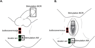
Nine patients underwent digital rectal examination (DRE) and sEMG, assessing voluntary anal contraction and sacral spinal reflexes (bulbocavernosus reflex and the anal wink). Our sEMG technique utilized surface recording electrodes and tactile elicitation of reflexes. EMG signal was acquired at bedside through the Noraxon MR3 system.
Results
It was quick, well accepted and did no harm. We found that contrary to the DRE, sEMG detected subclinical sacral motor activity and reflexes in 20% of cases for voluntary anal contraction and 40% of cases for the anal wink.
Conclusion
We believe our sEMG technique is a powerful tool able to enhance management of patients suffering from acute neurological impairments and requiring sacral function assessment.
中文翻译:
床旁肌电图用于临床评估适用于急性神经系统疾病住院患者的骶运动和反射活动:一项试点研究
学习规划
试点队列研究。
客观的
开发并在床边实施骶骨肌电图 (sEMG) 技术,以确定因急性神经系统疾病住院的患者的骶骨运动活动和反射是否正常。
环境
蒙特利尔圣心医院 (Hôpital du Sacré-Coeur de Montréal) 是一家加拿大一级大学创伤中心,专门从事 SCI 护理。
方法
九名患者接受了直肠指检 (DRE) 和 sEMG,评估自主肛门收缩和骶脊髓反射(球海绵体肌反射和肛门眨眼)。我们的表面肌电图技术利用表面记录电极和触觉诱发反射。通过 Noraxon MR3 系统在床边采集肌电图信号。
结果
它很快、被广泛接受并且没有造成任何伤害。我们发现,与 DRE 相反,sEMG 在 20% 的自愿肛门收缩病例和 40% 的肛门眨眼病例中检测到亚临床骶骨运动活动和反射。
结论
我们相信,我们的表面肌电图技术是一种强大的工具,能够加强对患有急性神经损伤并需要进行骶功能评估的患者的管理。


 京公网安备 11010802027423号