Spinal Cord Series and Cases ( IF 0.7 ) Pub Date : 2024-07-11 , DOI: 10.1038/s41394-024-00661-2 Rahul Sachdeva , Lauren Rietchel , Megan Lee , Dmitri Krassioukov-Enns , John Ditunno , Andrei V. Krassioukov
|
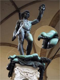
The Babinski sign is depicted in numerous historical artworks, with its earliest representation present 400 years before the formal description by Joseph Babinski [1,2,3]. This response is characterized by dorsiflexion of the hallux due to recruiting the extensor hallucis longus muscle, elicited by firmly stroking the foot from heel to toe with a blunt instrument. As a clinical diagnostic tool, the Babinski sign indicates injury to the pyramidal tracts, the primary motor pathway connecting cortical upper motor neurons (UMNs) with spinal cord motor neurons [also known as lower motor neurons (LMNs)]. This sign is present normally in infants, less than 24 months of age (due to incomplete myelination of the pyramidal tracts), and pathologically present in individuals who have sustained prolonged pyramidal tract dysfunction (e.g., stroke, spinal cord injury (SCI), multiple sclerosis and others) [4,5,6]. In elderly, gradually compressing the spinal cord and developing myelopathy from chronic spinal stenosis can produce the Babinski sign [7]. The Babinski sign was depicted in several medieval Madonna and Child paintings (e.g., Madonna and Child with Angels by Sandro Botticelli in 1468) [1]. All portray distinct hallux dorsiflexion expected in infants, demonstrating attempts of anatomical precision during this time.
After spinal cord injury, a specific pattern of reflex recovery occurs and can be classified into four distinct phases [4]. Immediately upon SCI, in phase 1 of spinal shock all reflexes are initially absent. A few hours after injury, pathological deep tendon reflexes may appear transiently. The Babinski sign can return as early as 1–3 days post-injury, during phase 2, if subjects are elderly [6]. Usually, the sign appears 4–30 days post-injury in phase 3, closely following deep tendon reflex recovery. In phase 4, the Babinski reflex becomes hyperactive. Overall, the sign develops after other reflexes appear indicating recovery from spinal shock. However, pre-existing injury or chronic damage to UMN pathways (e.g., stroke, myelopathy etc.) may cause the Babinski sign to be observed earlier [4]. The Babinski sign is probably the most notable reflex to return, with its manifestation positively predicting the presence of a pyramidal tract lesion [10].
中文翻译:
《珀尔修斯与美杜莎的头》中的巴宾斯基符号:通过神经学镜头重新审视艺术
巴宾斯基符号在许多历史艺术品中都有描绘,其最早的表现形式比约瑟夫·巴宾斯基 (Joseph Babinski) 的正式描述早 400 年 [1,2,3]。这种反应的特点是拇长背屈,这是由于用钝器从脚后跟到脚趾用力抚摸足部引起的拇长伸肌的募集所致。作为一种临床诊断工具,巴宾斯基征表明锥体束损伤,锥体束是连接皮质上运动神经元 (UMN) 与脊髓运动神经元 [也称为下运动神经元 (LMN)] 的主要运动通路。该体征通常出现在 24 个月以下的婴儿中(由于锥体束髓鞘形成不完全),并且病理性地出现在长期锥体束功能障碍(例如中风、脊髓损伤 (SCI)、多发性脊髓损伤)的个体中。硬化症等)[4,5,6]。在老年人中,慢性椎管狭窄逐渐压迫脊髓并发展为脊髓病,可产生巴宾斯基征[7]。巴宾斯基标志出现在几幅中世纪的《圣母子》画作中(例如桑德罗·波提切利于 1468 年创作的《圣母子与天使》)[1]。所有这些都描绘了婴儿所预期的明显的拇背屈,展示了这一时期解剖学精确性的尝试。
脊髓损伤后,会出现特定的反射恢复模式,可分为四个不同的阶段[4]。 SCI 发生后,在脊髓休克的第一阶段,所有反射最初都消失。受伤后数小时,可能会短暂出现病理性深部腱反射。如果受试者年龄较大,巴宾斯基征最早可在受伤后 1-3 天(即第 2 阶段)恢复 [6]。通常,该体征出现在受伤后 4-30 天的第 3 阶段,紧随深腱反射恢复之后。在第四阶段,巴宾斯基反射变得过度活跃。总体而言,该症状是在其他反射出现后出现的,表明脊髓休克已恢复。然而,先前存在的 UMN 通路损伤或慢性损伤(例如中风、脊髓病等)可能会导致更早观察到巴宾斯基征 [4]。巴宾斯基征可能是最显着的返回反射,其表现积极预测锥体束病变的存在[10]。


 京公网安备 11010802027423号