Nature Biomedical Engineering ( IF 26.8 ) Pub Date : 2024-07-10 , DOI: 10.1038/s41551-024-01217-3 Mustafa Nasir-Moin 1 , Lisa Irina Wadiura 2 , Vlad Sacalean 3, 4, 5 , Devin Juros 1 , Misha Movahed-Ezazi 6 , Emily K Lock 1 , Andrew Smith 1 , Matthew Lee 7 , Hannah Weiss 1 , Michael Müther 8 , Daniel Alber 1 , Sujay Ratna 9 , Camila Fang 6 , Eric Suero-Molina 8 , Sönke Hellwig 8 , Walter Stummer 8 , Karl Rössler 2 , Johannes A Hainfellner 10 , Georg Widhalm 2 , Barbara Kiesel 2 , David Reichert 11 , Mario Mischkulnig 2 , Rajan Jain 7 , Jakob Straehle 3, 4, 12 , Nicolas Neidert 3, 4, 12 , Oliver Schnell 3, 4, 13 , Jürgen Beck 3, 4, 14 , Jay Trautman 9 , Steve Pastore 9 , Donato Pacione 1 , Dimitris Placantonakis 1 , Eric Karl Oermann 1, 15 , John G Golfinos 1 , Todd C Hollon 16 , Matija Snuderl 6 , Christian W Freudiger 9 , Dieter Henrik Heiland 3, 4, 5, 17, 18 , Daniel A Orringer 1, 6
|
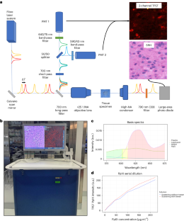
The most widely used fluorophore in glioma-resection surgery, 5-aminolevulinic acid (5-ALA), is thought to cause the selective accumulation of fluorescent protoporphyrin IX (PpIX) in tumour cells. Here we show that the clinical detection of PpIX can be improved via a microscope that performs paired stimulated Raman histology and two-photon excitation fluorescence microscopy (TPEF). We validated the technique in fresh tumour specimens from 115 patients with high-grade gliomas across four medical institutions. We found a weak negative correlation between tissue cellularity and the fluorescence intensity of PpIX across all imaged specimens. Semi-supervised clustering of the TPEF images revealed five distinct patterns of PpIX fluorescence, and spatial transcriptomic analyses of the imaged tissue showed that myeloid cells predominate in areas where PpIX accumulates in the intracellular space. Further analysis of external spatially resolved metabolomics, transcriptomics and RNA-sequencing datasets from glioblastoma specimens confirmed that myeloid cells preferentially accumulate and metabolize PpIX. Our findings question 5-ALA-induced fluorescence in glioma cells and show how 5-ALA and TPEF imaging can provide a window into the immune microenvironment of gliomas.
中文翻译:
通过配对刺激拉曼组织学和荧光显微镜在神经胶质瘤切除手术中定位原卟啉 IX
神经胶质瘤切除手术中使用最广泛的荧光团是 5-氨基乙酰丙酸 (5-ALA),被认为会导致荧光原卟啉 IX (PpIX) 在肿瘤细胞中选择性积累。在这里,我们表明,通过执行配对受激拉曼组织学和双光子激发荧光显微镜 (TPEF) 的显微镜可以改善 PpIX 的临床检测。我们在来自四家医疗机构的 115 名高级别神经胶质瘤患者的新鲜肿瘤标本中验证了该技术。我们发现所有成像样本中的组织细胞结构和 PpIX 荧光强度之间存在微弱的负相关性。 TPEF 图像的半监督聚类揭示了 PpIX 荧光的五种不同模式,成像组织的空间转录组分析表明,骨髓细胞在 PpIX 在细胞内空间积聚的区域中占主导地位。对胶质母细胞瘤标本的外部空间解析代谢组学、转录组学和 RNA 测序数据集的进一步分析证实,骨髓细胞优先积累和代谢 PpIX。我们的研究结果对神经胶质瘤细胞中 5-ALA 诱导的荧光提出了质疑,并展示了 5-ALA 和 TPEF 成像如何为了解神经胶质瘤的免疫微环境提供了一个窗口。


 京公网安备 11010802027423号