当前位置:
X-MOL 学术
›
Environ. Sci.: Nano
›
论文详情
Our official English website, www.x-mol.net, welcomes your
feedback! (Note: you will need to create a separate account there.)
Morphologic alterations across three levels of biological organization following oral exposure to silver-polymer nanocomposites in Japanese medaka (Oryzias latipes)
Environmental Science: Nano ( IF 5.8 ) Pub Date : 2024-07-03 , DOI: 10.1039/d4en00368c Melissa Chernick 1 , Alan J. Kennedy 2 , Treye Thomas 3 , Keana C. K. Scott 4 , Joana Marie Sipe 5 , Christine Ogilvie Hendren 5, 6 , Mark R. Wiesner 5 , David E. Hinton 1
Environmental Science: Nano ( IF 5.8 ) Pub Date : 2024-07-03 , DOI: 10.1039/d4en00368c Melissa Chernick 1 , Alan J. Kennedy 2 , Treye Thomas 3 , Keana C. K. Scott 4 , Joana Marie Sipe 5 , Christine Ogilvie Hendren 5, 6 , Mark R. Wiesner 5 , David E. Hinton 1
Affiliation

|
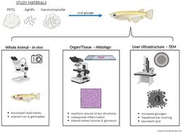
Polymer nanocomposites have diverse industrial and commercial uses. While many toxicity studies have assessed the individual materials (e.g., polymer, nanomaterial) comprising nanocomposites, few have examined the potential toxicity of the nanocomposite as a whole. Furthermore, products undergo machining during their manufacture and/or degradation as they age thereby resulting in potentially altered mixture exposures and differential effects compared to the parent nanocomposite. We assessed potential toxic effects of repeated oral exposure to silver nanoparticle (AgNP) embedded nanocomposites and compared them to individual component materials using a transparent strain of the fish model Japanese medaka (Oryzias latipes). This strain allowed for the comparison of morphologic alterations at three levels of biological organization: whole animal; organ/tissue by examination of histologic sections; and, subcellular using transmission electron microscopy (TEM). Adult fish were exposed to AgNPs, silver nitrate, abraded PETG microplastics, or abraded nanocomposites via 7 oral gavages over the course of 2 weeks. In vivo observations showed alterations in the liver and gallbladder of fish exposed to pristine AgNPs and nanocomposites. When histologic sections of these same individuals were examined by light microscopy, hepatic and biliary alterations were observed. Similarly, alterations of the head kidney were also observed in fish exposed to silver and its composites, with both tubules and glomeruli affected. In both of these organs, many of the changes occurred adjacent to large blood vessels, suggesting material translocated from the bloodstream to adjacent tissues. TEM of the liver supported histological findings, with increased glycogen as well as hepatocellular swelling and abundant lipid vesicles in exposure groups. Very few morphological changes at any level of biological organization were observed in fish exposed to the plastic matrix alone. This transparent fish model proved advantageous in evaluating risks and potential human health concerns associated with the ingestion of silver nanocomposites.
中文翻译:

日本青鳉(Oryzias latipes)口服银聚合物纳米复合材料后生物组织三个层面的形态变化
聚合物纳米复合材料具有多种工业和商业用途。虽然许多毒性研究评估了包含纳米复合材料的单个材料(例如聚合物、纳米材料),但很少有人研究纳米复合材料作为一个整体的潜在毒性。此外,产品在制造过程中会经历机械加工和/或随着老化而降解,从而导致与母体纳米复合材料相比可能改变混合物暴露和差异效应。我们评估了反复口服暴露于嵌入银纳米粒子 (AgNP) 的纳米复合材料的潜在毒性作用,并使用日本青鳉鱼模型 (Oryzias latipes) 的透明品系将其与单独的成分材料进行比较。该菌株允许比较生物组织三个层面的形态变化:整个动物;通过组织切片检查来检查器官/组织;使用透射电子显微镜 (TEM) 进行亚细胞观察。在 2 周内,通过 7 次口服管饲法,将成年鱼暴露于 AgNP、硝酸银、磨损的 PETG 微塑料或磨损的纳米复合材料中。体内观察显示,暴露于原始银纳米粒子和纳米复合材料的鱼的肝脏和胆囊发生了变化。当通过光学显微镜检查这些相同个体的组织学切片时,观察到肝脏和胆道的改变。同样,在接触银及其复合物的鱼中也观察到头肾的改变,肾小管和肾小球都受到影响。在这两个器官中,许多变化发生在大血管附近,表明物质从血流转移到邻近组织。 肝脏的 TEM 支持组织学结果,暴露组中糖原增加以及肝细胞肿胀和丰富的脂质囊泡。在仅暴露于塑料基质的鱼中,在任何级别的生物组织中都观察到很少的形态变化。事实证明,这种透明的鱼模型在评估与摄入银纳米复合材料相关的风险和潜在人类健康问题方面具有优势。
更新日期:2024-07-03
中文翻译:

日本青鳉(Oryzias latipes)口服银聚合物纳米复合材料后生物组织三个层面的形态变化
聚合物纳米复合材料具有多种工业和商业用途。虽然许多毒性研究评估了包含纳米复合材料的单个材料(例如聚合物、纳米材料),但很少有人研究纳米复合材料作为一个整体的潜在毒性。此外,产品在制造过程中会经历机械加工和/或随着老化而降解,从而导致与母体纳米复合材料相比可能改变混合物暴露和差异效应。我们评估了反复口服暴露于嵌入银纳米粒子 (AgNP) 的纳米复合材料的潜在毒性作用,并使用日本青鳉鱼模型 (Oryzias latipes) 的透明品系将其与单独的成分材料进行比较。该菌株允许比较生物组织三个层面的形态变化:整个动物;通过组织切片检查来检查器官/组织;使用透射电子显微镜 (TEM) 进行亚细胞观察。在 2 周内,通过 7 次口服管饲法,将成年鱼暴露于 AgNP、硝酸银、磨损的 PETG 微塑料或磨损的纳米复合材料中。体内观察显示,暴露于原始银纳米粒子和纳米复合材料的鱼的肝脏和胆囊发生了变化。当通过光学显微镜检查这些相同个体的组织学切片时,观察到肝脏和胆道的改变。同样,在接触银及其复合物的鱼中也观察到头肾的改变,肾小管和肾小球都受到影响。在这两个器官中,许多变化发生在大血管附近,表明物质从血流转移到邻近组织。 肝脏的 TEM 支持组织学结果,暴露组中糖原增加以及肝细胞肿胀和丰富的脂质囊泡。在仅暴露于塑料基质的鱼中,在任何级别的生物组织中都观察到很少的形态变化。事实证明,这种透明的鱼模型在评估与摄入银纳米复合材料相关的风险和潜在人类健康问题方面具有优势。


 京公网安备 11010802027423号