当前位置:
X-MOL 学术
›
Anal. Chim. Acta
›
论文详情
Our official English website, www.x-mol.net, welcomes your feedback! (Note: you will need to create a separate account there.)
Unveiling compositional images of specific proteins in individual cells by LA-ICP-MS: Labelling with ruthenium red and metal nanoclusters
Analytica Chimica Acta ( IF 5.7 ) Pub Date : 2024-06-25 , DOI: 10.1016/j.aca.2024.342906 Paula Menero-Valdés , Lydia Álvarez , Héctor González-Iglesias , Beatriz Fernández , Rosario Pereiro
Analytica Chimica Acta ( IF 5.7 ) Pub Date : 2024-06-25 , DOI: 10.1016/j.aca.2024.342906 Paula Menero-Valdés , Lydia Álvarez , Héctor González-Iglesias , Beatriz Fernández , Rosario Pereiro
|
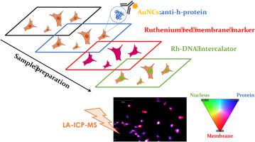
Recent biological studies have demonstrated that changes can occur in the cellular genome and proteome due to variations in cell volume. Therefore, it is imperative to take cell volume into account when analyzing a target protein. This consideration becomes especially critical in experimental models involving cells subjected to different treatments. Failure to consider cell volume could obscure the studied biological phenomena or lead to erroneous conclusions. However, quantitative imaging of proteins within cells by LA-ICP-MS is limited by the lack of methods that provide the protein concentration (protein mass over cell volume) rather than just protein mass within individual cells. The combination of a metal tagged immunoprobe with ruthenium red (RR) labelling enables the simultaneous analysis of a specific protein and the cell volume in each cell analyzed by LA-ICP-(Q)MS. The results indicate that the CYP1B1 concentration exhibits a quasi–normally distribution in control ARPE-19 cells, whereas AAPH–treated cells reveal the presence of two distinct cell groups, responding and non–responding cells to an induced oxidative stress. The labelling of the membrane with RR and the measurement of Ru mass in each cell by LA-ICP-MS offers higher precision compared to manually delimitation of the cell perimeter and eliminates the risk of biased information, which can be prone to inter–observer variability. The proposed procedure is fast and minimizes errors in cell area assignment and offers the possibility to carry out a faster data treatment approach if just relative volumes are compared, which can be advantageous for specific applications. This work presents an innovative strategy to directly study the distribution and concentration of proteins within individual cells by LA-ICP-MS. This method employs ruthenium red as a cell volume marker and Au nanoclusters (AuNCs) tagged immunoprobes to label the protein of interest. Furthermore, the proposed labelling strategy enables rapid data processing, allowing for the calculation of relative concentrations and thus facilitating the comparison across large datasets. As a proof–of–concept, the concentration of the CYP1B1 protein was quantified in ARPE-19 cells under both control and oxidative stress conditions.
中文翻译:

通过 LA-ICP-MS 揭示单个细胞中特定蛋白质的成分图像:用钌红和金属纳米团簇进行标记
最近的生物学研究表明,由于细胞体积的变化,细胞基因组和蛋白质组可能发生变化。因此,在分析目标蛋白时必须考虑细胞体积。这种考虑在涉及接受不同处理的细胞的实验模型中变得尤其重要。不考虑细胞体积可能会掩盖所研究的生物现象或导致错误的结论。然而,通过 LA-ICP-MS 对细胞内蛋白质进行定量成像受到限制,因为缺乏提供蛋白质浓度(蛋白质质量除以细胞体积)而不仅仅是单个细胞内蛋白质质量的方法。金属标记免疫探针与钌红 (RR) 标记相结合,可以同时分析特定蛋白质和通过 LA-ICP-(Q)MS 分析的每个细胞中的细胞体积。结果表明,对照 ARPE-19 细胞中的 CYP1B1 浓度呈现准正态分布,而 AAPH 处理的细胞则显示出两个不同的细胞组的存在,即对诱导的氧化应激有反应的细胞和无反应的细胞。与手动划定细胞周长相比,用 RR 标记膜并通过 LA-ICP-MS 测量每个细胞中的 Ru 质量可提供更高的精度,并消除容易出现观察者间差异的信息偏差风险。所提出的程序速度快,并且最大限度地减少了单元区域分配中的错误,并且如果仅比较相对体积,则提供了执行更快的数据处理方法的可能性,这对于特定应用来说是有利的。 这项工作提出了一种创新策略,通过 LA-ICP-MS 直接研究单个细胞内蛋白质的分布和浓度。该方法采用钌红作为细胞体积标记,并使用金纳米簇 (AuNC) 标记的免疫探针来标记感兴趣的蛋白质。此外,所提出的标记策略可以实现快速数据处理,允许计算相对浓度,从而促进大型数据集之间的比较。作为概念验证,我们在对照和氧化应激条件下对 ARPE-19 细胞中 CYP1B1 蛋白的浓度进行了定量。
更新日期:2024-06-25
中文翻译:
通过 LA-ICP-MS 揭示单个细胞中特定蛋白质的成分图像:用钌红和金属纳米团簇进行标记
最近的生物学研究表明,由于细胞体积的变化,细胞基因组和蛋白质组可能发生变化。因此,在分析目标蛋白时必须考虑细胞体积。这种考虑在涉及接受不同处理的细胞的实验模型中变得尤其重要。不考虑细胞体积可能会掩盖所研究的生物现象或导致错误的结论。然而,通过 LA-ICP-MS 对细胞内蛋白质进行定量成像受到限制,因为缺乏提供蛋白质浓度(蛋白质质量除以细胞体积)而不仅仅是单个细胞内蛋白质质量的方法。金属标记免疫探针与钌红 (RR) 标记相结合,可以同时分析特定蛋白质和通过 LA-ICP-(Q)MS 分析的每个细胞中的细胞体积。结果表明,对照 ARPE-19 细胞中的 CYP1B1 浓度呈现准正态分布,而 AAPH 处理的细胞则显示出两个不同的细胞组的存在,即对诱导的氧化应激有反应的细胞和无反应的细胞。与手动划定细胞周长相比,用 RR 标记膜并通过 LA-ICP-MS 测量每个细胞中的 Ru 质量可提供更高的精度,并消除容易出现观察者间差异的信息偏差风险。所提出的程序速度快,并且最大限度地减少了单元区域分配中的错误,并且如果仅比较相对体积,则提供了执行更快的数据处理方法的可能性,这对于特定应用来说是有利的。 这项工作提出了一种创新策略,通过 LA-ICP-MS 直接研究单个细胞内蛋白质的分布和浓度。该方法采用钌红作为细胞体积标记,并使用金纳米簇 (AuNC) 标记的免疫探针来标记感兴趣的蛋白质。此外,所提出的标记策略可以实现快速数据处理,允许计算相对浓度,从而促进大型数据集之间的比较。作为概念验证,我们在对照和氧化应激条件下对 ARPE-19 细胞中 CYP1B1 蛋白的浓度进行了定量。


 京公网安备 11010802027423号