当前位置:
X-MOL 学术
›
Redox Biol.
›
论文详情
Our official English website, www.x-mol.net, welcomes your feedback! (Note: you will need to create a separate account there.)
NADPH Alters DUOX1 Calcium Responsiveness
Redox Biology ( IF 10.7 ) Pub Date : 2024-06-20 , DOI: 10.1016/j.redox.2024.103251 Gregory E. Conner
Redox Biology ( IF 10.7 ) Pub Date : 2024-06-20 , DOI: 10.1016/j.redox.2024.103251 Gregory E. Conner
|
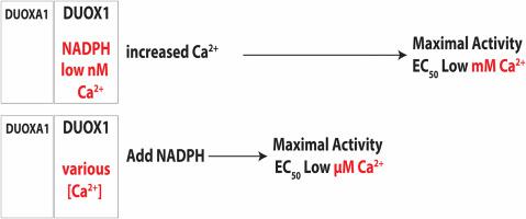
Hydrogen peroxide is a key element in redox signaling and in setting cellular redox tone. DUOX1 and DUOX2, that directly synthesize hydrogen peroxide, are the most abundant NADPH oxidase transcripts in most epithelia. DUOX1 and DUOX2 hydrogen peroxide synthesis is regulated by intracellular calcium transients and thus cells can respond to signals and initiate responses by increasing cellular hydrogen peroxide synthesis. Nevertheless, many details of their enzymatic regulation are still unexplored. DUOX1 and DUOXA1 were expressed in HEK293T cells and activity was studied in homogenates and membrane fractions. When DUOX1 homogenates or membranes were pre-incubated in NADPH and started with addition of Ca, to mimic intracellular activation, progress curves were distinctly different from those pre-incubated in Ca and started with NADPH. The Ca EC for DUOX1's initial rate when pre-incubated in Ca, was three orders of magnitude lower (EC ∼ 10 M) than with preincubation in NADPH (EC ∼ 10 M). In addition, activity was several fold lower with Ca start. Identical results were obtained using homogenates and membrane fractions. The data suggested that DUOX1 Ca binding in expected physiological signaling conditions only slowly leads to maximal hydrogen peroxide synthesis and that full hydrogen peroxide synthesis activity only can occur when encountering extremely high concentration Ca signals. Thus, a complex interplay of intracellular NADPH and Ca concentrations regulate DUOX1 over a wide extent and may limit DUOX1 activity to a restricted range and spatial distribution.
中文翻译:

NADPH 改变 DUOX1 钙反应
过氧化氢是氧化还原信号传导和设定细胞氧化还原基调的关键元素。 DUOX1 和 DUOX2 直接合成过氧化氢,是大多数上皮细胞中最丰富的 NADPH 氧化酶转录本。 DUOX1 和 DUOX2 过氧化氢合成受细胞内钙瞬变调节,因此细胞可以响应信号并通过增加细胞过氧化氢合成来启动反应。然而,它们的酶调节的许多细节仍有待探索。 DUOX1 和 DUOXA1 在 HEK293T 细胞中表达,并在匀浆和膜组分中研究活性。当 DUOX1 匀浆或膜在 NADPH 中预孵育并开始添加 Ca 来模拟细胞内激活时,进度曲线与在 Ca 中预孵育并从 NADPH 开始的曲线明显不同。在 Ca 中预孵育时,DUOX1 的 Ca EC 初始速率 (EC ~ 10 M) 比在 NADPH 中预孵育 (EC ~ 10 M) 低三个数量级。此外,Ca 启动后的活性降低了几倍。使用匀浆和膜组分获得了相同的结果。数据表明,在预期的生理信号条件下,DUOX1 Ca 结合只会缓慢地导致最大的过氧化氢合成,并且只有在遇到极高浓度的 Ca 信号时,才会发生完全的过氧化氢合成活性。因此,细胞内 NADPH 和 Ca 浓度的复杂相互作用在很大程度上调节 DUOX1,并可能将 DUOX1 活性限制在有限的范围和空间分布内。
更新日期:2024-06-20
中文翻译:
NADPH 改变 DUOX1 钙反应
过氧化氢是氧化还原信号传导和设定细胞氧化还原基调的关键元素。 DUOX1 和 DUOX2 直接合成过氧化氢,是大多数上皮细胞中最丰富的 NADPH 氧化酶转录本。 DUOX1 和 DUOX2 过氧化氢合成受细胞内钙瞬变调节,因此细胞可以响应信号并通过增加细胞过氧化氢合成来启动反应。然而,它们的酶调节的许多细节仍有待探索。 DUOX1 和 DUOXA1 在 HEK293T 细胞中表达,并在匀浆和膜组分中研究活性。当 DUOX1 匀浆或膜在 NADPH 中预孵育并开始添加 Ca 来模拟细胞内激活时,进度曲线与在 Ca 中预孵育并从 NADPH 开始的曲线明显不同。在 Ca 中预孵育时,DUOX1 的 Ca EC 初始速率 (EC ~ 10 M) 比在 NADPH 中预孵育 (EC ~ 10 M) 低三个数量级。此外,Ca 启动后的活性降低了几倍。使用匀浆和膜组分获得了相同的结果。数据表明,在预期的生理信号条件下,DUOX1 Ca 结合只会缓慢地导致最大的过氧化氢合成,并且只有在遇到极高浓度的 Ca 信号时,才会发生完全的过氧化氢合成活性。因此,细胞内 NADPH 和 Ca 浓度的复杂相互作用在很大程度上调节 DUOX1,并可能将 DUOX1 活性限制在有限的范围和空间分布内。


 京公网安备 11010802027423号