Cell ( IF 45.5 ) Pub Date : 2024-06-24 , DOI: 10.1016/j.cell.2024.05.055 Marie Schott , Daniel León-Periñán , Elena Splendiani , Leon Strenger , Jan Robin Licha , Tancredi Massimo Pentimalli , Simon Schallenberg , Jonathan Alles , Sarah Samut Tagliaferro , Anastasiya Boltengagen , Sebastian Ehrig , Stefano Abbiati , Steffen Dommerich , Massimiliano Pagani , Elisabetta Ferretti , Giuseppe Macino , Nikos Karaiskos , Nikolaus Rajewsky
|
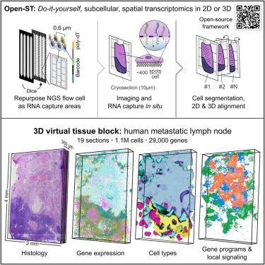
Spatial transcriptomics (ST) methods unlock molecular mechanisms underlying tissue development, homeostasis, or disease. However, there is a need for easy-to-use, high-resolution, cost-efficient, and 3D-scalable methods. Here, we report Open-ST, a sequencing-based, open-source experimental and computational resource to address these challenges and to study the molecular organization of tissues in 2D and 3D. In mouse brain, Open-ST captured transcripts at subcellular resolution and reconstructed cell types. In primary head-and-neck tumors and patient-matched healthy/metastatic lymph nodes, Open-ST captured the diversity of immune, stromal, and tumor populations in space, validated by imaging-based ST. Distinct cell states were organized around cell-cell communication hotspots in the tumor but not the metastasis. Strikingly, the 3D reconstruction and multimodal analysis of the metastatic lymph node revealed spatially contiguous structures not visible in 2D and potential biomarkers precisely at the 3D tumor/lymph node boundary. All protocols and software are available at https://rajewsky-lab.github.io/openst.
中文翻译:
Open-ST:3D 高分辨率空间转录组学
空间转录组学 (ST) 方法揭示了组织发育、稳态或疾病的分子机制。然而,需要易于使用、高分辨率、经济高效且 3D 可扩展的方法。在此,我们报告了 Open-ST,这是一种基于测序的开源实验和计算资源,旨在解决这些挑战并研究 2D 和 3D 组织的分子组织。在小鼠大脑中,Open-ST 以亚细胞分辨率捕获转录本并重建细胞类型。在原发性头颈肿瘤和患者匹配的健康/转移淋巴结中,Open-ST 捕获了空间中免疫、间质和肿瘤群体的多样性,并通过基于成像的 ST 进行了验证。不同的细胞状态是围绕肿瘤中的细胞间通讯热点组织的,而不是转移灶。引人注目的是,转移淋巴结的 3D 重建和多模态分析揭示了 2D 中不可见的空间连续结构以及精确位于 3D 肿瘤/淋巴结边界的潜在生物标志物。所有协议和软件均可在 https://rajewsky-lab.github.io/openst 上获取。


 京公网安备 11010802027423号