当前位置:
X-MOL 学术
›
Anal. Chim. Acta
›
论文详情
Our official English website, www.x-mol.net, welcomes your feedback! (Note: you will need to create a separate account there.)
One-step detection of procollagen type III N-terminal peptide as a fibrosis biomarker using fluorescent immunosensor (quenchbody)
Analytica Chimica Acta ( IF 5.7 ) Pub Date : 2024-06-18 , DOI: 10.1016/j.aca.2024.342887 Joon-Yeop Yi , Jaewon Ryu , Yujin Jeong , Yoeseph Cho , Minyoung Kim , Mijin Jeon , Hee Ho Park , Nathaniel S. Hwang , Hee-Jin Jeong , Changmin Sung
Analytica Chimica Acta ( IF 5.7 ) Pub Date : 2024-06-18 , DOI: 10.1016/j.aca.2024.342887 Joon-Yeop Yi , Jaewon Ryu , Yujin Jeong , Yoeseph Cho , Minyoung Kim , Mijin Jeon , Hee Ho Park , Nathaniel S. Hwang , Hee-Jin Jeong , Changmin Sung
|
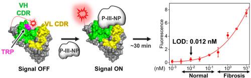
Procollagen type III N-terminal peptide (P-III-NP) is a fibrosis biomarker associated with liver and cardiac fibrosis. Despite the value of P-III-NP as a biomarker, its analysis currently relies on enzyme-linked immunosorbent assays (ELISA) and radioimmunoassays (RIA), which require more than 3 h. To facilitate early diagnosis and treatment through rapid biomarker testing, we developed a one-step immunoassay for P-III-NP using a quenchbody, which is a fluorescence-labeled immunosensor for immediate signal generation. To create quenchbodies, the total mRNA of P-III-NP antibodies was extracted from early-developed hybridoma cells, and genes of variable regions were obtained through cDNA synthesis, inverse PCR, and sequencing. A single-chain variable fragment (scFv) with an N-terminal Cys-tag was expressed in Shuffle T7, resulting in a final yield of 9.8 mg L. The fluorescent dye was labeled on the Cys-tag of the anti-P-III-NP scFv using maleimide-thiol click chemistry, and the spacer arm lengths between the maleimide-fluorescent dyes were compared. Consequently, a TAMRA-C-labeled quenchbody exhibited antigen-dependent fluorescence signals and demonstrated its ability to detect P-III-NP at concentrations as low as 0.46 ng mL for buffer samples, 1.0 ng mL for 2 % human serum samples. This one-step P-III-NP detection method provides both qualitative and quantitative outcomes within a concise 5-min timeframe. Furthermore, its application can be expanded using a 96-well platform and human serum, making it a high-throughput and sensitive method for testing fibrotic biomarkers.
中文翻译:

使用荧光免疫传感器(猝灭体)一步检测 III 型前胶原 N 端肽作为纤维化生物标志物
III 型原胶原 N 端肽 (P-III-NP) 是一种与肝脏和心脏纤维化相关的纤维化生物标志物。尽管 P-III-NP 作为生物标志物具有价值,但其分析目前依赖于酶联免疫吸附测定 (ELISA) 和放射免疫测定 (RIA),这需要超过 3 小时。为了通过快速生物标志物测试促进早期诊断和治疗,我们开发了一种使用猝灭体的 P-III-NP 一步式免疫测定法,猝灭体是一种用于立即产生信号的荧光标记免疫传感器。为了创建猝灭体,从早期发育的杂交瘤细胞中提取P-III-NP抗体的总mRNA,并通过cDNA合成、反向PCR和测序获得可变区基因。带有 N 端 Cys 标签的单链可变片段 (scFv) 在 Shuffle T7 中表达,最终产量为 9.8 mg/L。荧光染料标记在抗 P-III 的 Cys 标签上-NP scFv 使用马来酰亚胺-硫醇点击化学,并比较马来酰亚胺-荧光染料之间的间隔臂长度。因此,TAMRA-C 标记的猝灭体表现出抗原依赖性荧光信号,并证明其能够在缓冲液样品中检测浓度低至 0.46 ng mL 的 P-III-NP,在 2% 人血清样品中检测浓度低至 1.0 ng mL 的 P-III-NP。这种一步式 P-III-NP 检测方法可在简洁的 5 分钟时间内提供定性和定量结果。此外,它的应用可以使用 96 孔平台和人血清进行扩展,使其成为测试纤维化生物标志物的高通量和灵敏方法。
更新日期:2024-06-18
中文翻译:
使用荧光免疫传感器(猝灭体)一步检测 III 型前胶原 N 端肽作为纤维化生物标志物
III 型原胶原 N 端肽 (P-III-NP) 是一种与肝脏和心脏纤维化相关的纤维化生物标志物。尽管 P-III-NP 作为生物标志物具有价值,但其分析目前依赖于酶联免疫吸附测定 (ELISA) 和放射免疫测定 (RIA),这需要超过 3 小时。为了通过快速生物标志物测试促进早期诊断和治疗,我们开发了一种使用猝灭体的 P-III-NP 一步式免疫测定法,猝灭体是一种用于立即产生信号的荧光标记免疫传感器。为了创建猝灭体,从早期发育的杂交瘤细胞中提取P-III-NP抗体的总mRNA,并通过cDNA合成、反向PCR和测序获得可变区基因。带有 N 端 Cys 标签的单链可变片段 (scFv) 在 Shuffle T7 中表达,最终产量为 9.8 mg/L。荧光染料标记在抗 P-III 的 Cys 标签上-NP scFv 使用马来酰亚胺-硫醇点击化学,并比较马来酰亚胺-荧光染料之间的间隔臂长度。因此,TAMRA-C 标记的猝灭体表现出抗原依赖性荧光信号,并证明其能够在缓冲液样品中检测浓度低至 0.46 ng mL 的 P-III-NP,在 2% 人血清样品中检测浓度低至 1.0 ng mL 的 P-III-NP。这种一步式 P-III-NP 检测方法可在简洁的 5 分钟时间内提供定性和定量结果。此外,它的应用可以使用 96 孔平台和人血清进行扩展,使其成为测试纤维化生物标志物的高通量和灵敏方法。


 京公网安备 11010802027423号