Nature ( IF 50.5 ) Pub Date : 2024-06-19 , DOI: 10.1038/s41586-024-07563-1 Katherine Benjamin 1 , Aneesha Bhandari 2, 3 , Jessica D Kepple 2, 3 , Rui Qi 2, 3, 4 , Zhouchun Shang 5, 6 , Yanan Xing 5, 6 , Yanru An 5 , Nannan Zhang 7 , Yong Hou 5 , Tanya L Crockford 2, 3 , Oliver McCallion 8 , Fadi Issa 8 , Joanna Hester 8 , Ulrike Tillmann 1, 9 , Heather A Harrington 1, 2, 10, 11, 12 , Katherine R Bull 2, 3, 4
|
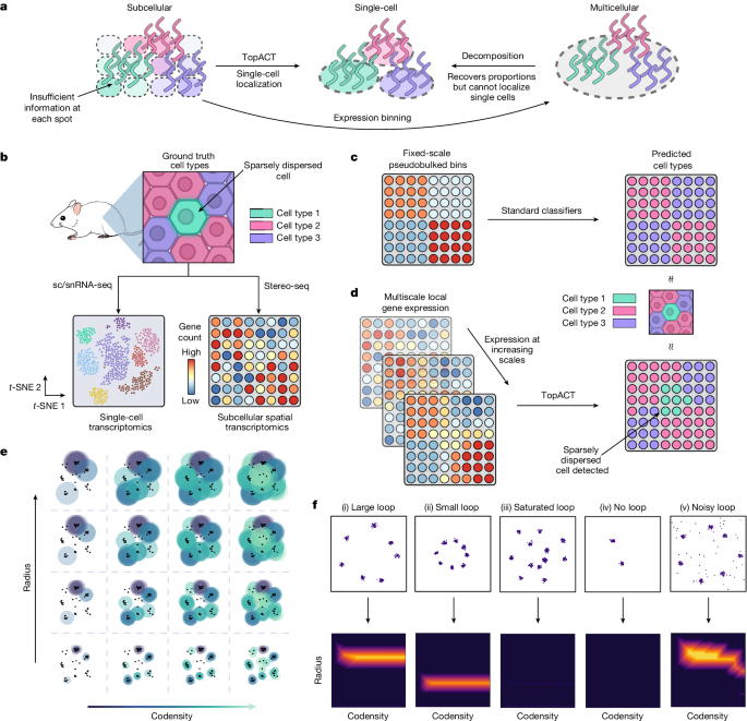
Spatial transcriptomics measures in situ gene expression at millions of locations within a tissue1, hitherto with some trade-off between transcriptome depth, spatial resolution and sample size2. Although integration of image-based segmentation has enabled impactful work in this context, it is limited by imaging quality and tissue heterogeneity. By contrast, recent array-based technologies offer the ability to measure the entire transcriptome at subcellular resolution across large samples3,4,5,6. Presently, there exist no approaches for cell type identification that directly leverage this information to annotate individual cells. Here we propose a multiscale approach to automatically classify cell types at this subcellular level, using both transcriptomic information and spatial context. We showcase this on both targeted and whole-transcriptome spatial platforms, improving cell classification and morphology for human kidney tissue and pinpointing individual sparsely distributed renal mouse immune cells without reliance on image data. By integrating these predictions into a topological pipeline based on multiparameter persistent homology7,8,9, we identify cell spatial relationships characteristic of a mouse model of lupus nephritis, which we validate experimentally by immunofluorescence. The proposed framework readily generalizes to new platforms, providing a comprehensive pipeline bridging different levels of biological organization from genes through to tissues.
中文翻译:
多尺度拓扑对亚细胞空间转录组学中的细胞进行分类
空间转录组学测量组织内数百万个位置的原位基因表达1 ,迄今为止在转录组深度、空间分辨率和样本大小2之间进行了一些权衡。尽管基于图像的分割的集成在这方面实现了有影响力的工作,但它受到成像质量和组织异质性的限制。相比之下,最近基于阵列的技术能够以亚细胞分辨率测量大样本的整个转录组3,4,5,6 。目前,不存在直接利用该信息来注释单个细胞的细胞类型识别方法。在这里,我们提出了一种多尺度方法,利用转录组信息和空间背景在亚细胞水平上自动分类细胞类型。我们在靶向和全转录组空间平台上展示了这一点,改进了人类肾脏组织的细胞分类和形态,并在不依赖图像数据的情况下精确定位单个稀疏分布的肾脏小鼠免疫细胞。通过将这些预测整合到基于多参数持久同源性的拓扑管道中7,8,9 ,我们确定了狼疮肾炎小鼠模型的细胞空间关系特征,并通过免疫荧光进行实验验证。所提出的框架很容易推广到新平台,提供一个全面的管道,连接从基因到组织的不同级别的生物组织。


 京公网安备 11010802027423号