Our official English website, www.x-mol.net, welcomes your
feedback! (Note: you will need to create a separate account there.)
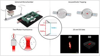
Two-photon microscopy of acoustofluidic trapping for highly sensitive cell analysis
Lab on a Chip ( IF 6.1 ) Pub Date : 2024-06-19 , DOI: 10.1039/d4lc00144c Thomas Kellerer 1 , Bettina Sailer 2 , Patrick Byers 1 , Rune Barnkob 3 , Oliver Hayden 2 , Thomas Hellerer 1
Lab on a Chip ( IF 6.1 ) Pub Date : 2024-06-19 , DOI: 10.1039/d4lc00144c Thomas Kellerer 1 , Bettina Sailer 2 , Patrick Byers 1 , Rune Barnkob 3 , Oliver Hayden 2 , Thomas Hellerer 1
Affiliation
|
We combine two-photon-excited fluorescence microscopy and acoustofluidic trapping in a spherical microchamber to in vitro study cells and cell clusters three-dimensionally close to in vivo conditions. The two-photon microscopy provides the in-depth 3D analysis of the spherical microchamber dimensions as well as the positions of trapped samples therein with high spatial precision and high temporal resolution enabling even tracking of the fast moving particles. Furthermore, optical sectioning allows to gather information of individual cells in trapped cell clusters inside the chamber. We demonstrate real-time monitoring of osmosis in A549 lung cells and red blood cells as one possible biomedical application. The observed osmosis reduced the cell membrane diameter by approximately 4 μm in the A549 cells and by approximately 2 μm in the red blood cells. Our approach provides an important optical tool for future investigations of cell functions and cell–cell interactions avoiding wall-contact inside an acoustofluidic device.
中文翻译:

用于高灵敏度细胞分析的声流捕获双光子显微镜
我们将双光子激发荧光显微镜和球形微室中的声流捕获相结合,在体外研究细胞和细胞簇的三维接近体内条件。双光子显微镜可对球形微室尺寸以及其中捕获的样品的位置进行深入的 3D 分析,具有高空间精度和高时间分辨率,甚至可以跟踪快速移动的粒子。此外,光学切片可以收集室内捕获细胞簇中单个细胞的信息。我们展示了 A549 肺细胞和红细胞渗透压的实时监测作为一种可能的生物医学应用。观察到的渗透作用使 A549 细胞的细胞膜直径减小了约 4 μm,使红细胞的细胞膜直径减小了约 2 μm。我们的方法为未来研究细胞功能和细胞间相互作用提供了重要的光学工具,避免声流体装置内的壁接触。
更新日期:2024-06-19
中文翻译:
用于高灵敏度细胞分析的声流捕获双光子显微镜
我们将双光子激发荧光显微镜和球形微室中的声流捕获相结合,在体外研究细胞和细胞簇的三维接近体内条件。双光子显微镜可对球形微室尺寸以及其中捕获的样品的位置进行深入的 3D 分析,具有高空间精度和高时间分辨率,甚至可以跟踪快速移动的粒子。此外,光学切片可以收集室内捕获细胞簇中单个细胞的信息。我们展示了 A549 肺细胞和红细胞渗透压的实时监测作为一种可能的生物医学应用。观察到的渗透作用使 A549 细胞的细胞膜直径减小了约 4 μm,使红细胞的细胞膜直径减小了约 2 μm。我们的方法为未来研究细胞功能和细胞间相互作用提供了重要的光学工具,避免声流体装置内的壁接触。


 京公网安备 11010802027423号