当前位置:
X-MOL 学术
›
Phys. Rev. X
›
论文详情
Our official English website, www.x-mol.net, welcomes your
feedback! (Note: you will need to create a separate account there.)
In Situ Magnetometry of Iron in Human Dopaminergic Neurons Using Superresolution MRI and Ion-Beam Microscopy
Physical Review X ( IF 11.6 ) Pub Date : 2024-06-10 , DOI: 10.1103/physrevx.14.021041 Malte Brammerloh 1, 2, 3 , Renat Sibgatulin 4 , Karl-Heinz Herrmann 4 , Markus Morawski 1, 3 , Tilo Reinert 1, 3 , Carsten Jäger 1, 3 , Roland Müller 1 , Gerald Falkenberg 5 , Dennis Brückner 5 , Kerrin J. Pine 1 , Andreas Deistung 6 , Valerij G. Kiselev 7 , Jürgen R. Reichenbach 4 , Nikolaus Weiskopf 1, 3, 8 , Evgeniya Kirilina 1
Physical Review X ( IF 11.6 ) Pub Date : 2024-06-10 , DOI: 10.1103/physrevx.14.021041 Malte Brammerloh 1, 2, 3 , Renat Sibgatulin 4 , Karl-Heinz Herrmann 4 , Markus Morawski 1, 3 , Tilo Reinert 1, 3 , Carsten Jäger 1, 3 , Roland Müller 1 , Gerald Falkenberg 5 , Dennis Brückner 5 , Kerrin J. Pine 1 , Andreas Deistung 6 , Valerij G. Kiselev 7 , Jürgen R. Reichenbach 4 , Nikolaus Weiskopf 1, 3, 8 , Evgeniya Kirilina 1
Affiliation
|
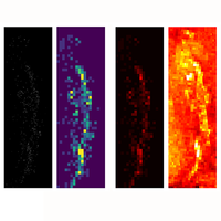
Paramagnetic transition metals play a crucial role as cofactors in various cellular catalytic processes. However, their high concentrations in reactive oxidation states can induce oxidative stress, resulting in cell dysfunction or death. Hence, it is vital to have methods to monitor metal concentrations and paramagnetic properties in cells for medicine and cell biology. Here we present a novel multimodal method for in-cell magnetometry enabling direct measurement of metal magnetic properties within individual cells in tissue, without prior isolation and at room temperature. Individual cell magnetic moments are measured using superresolution magnetic resonance imaging (MRI) microscopy at 9.4 T by detecting microscopic magnetic-field perturbations around the cells. The cellular metal content is quantified using ion-beam microscopy or synchrotron micro-x-ray fluorescence for the same cells. The metal magnetic susceptibility at 9.4 T is then obtained from the slope of the cell magnetic moments’ dependence on cell metal content. To estimate the susceptibility at lower fields, multifield MR relaxometry and biophysical modeling are employed, extrapolating the 9.4-T susceptibility values to fields as low as 3 T. We apply the new method to determine the susceptibility of iron accumulated in human dopaminergic neurons inside neuromelanin, the by-product of dopamine synthesis. The susceptibility of iron in neuromelanin is measured to be providing unique insights into the biochemistry of iron inside dopaminergic neurons. The obtained value reveals a predominant monoatomic low-affinity iron-binding site within neuromelanin, indicating a higher neurotoxicity of iron than previously suggested. Furthermore, the measured susceptibility value establishes a quantitative relationship between cellular iron concentration and iron-sensitive MRI parameters, which can be noninvasively measured in vivo. This breakthrough paves the way for the in vivo detection of dopaminergic neuron density and iron load, requiring a standard clinical MRI scanner only. It promises to facilitate early diagnosis of Parkinson’s disease. In conclusion, our presented novel method enables the direct measurements of magnetic properties of paramagnetic metals within single cells with high sensitivity and across large cell groups within a macroscopic volume, providing invaluable information about the cellular biology of metals.
中文翻译:

使用超分辨率 MRI 和离子束显微镜对人类多巴胺能神经元中的铁进行原位磁力测量
顺磁性过渡金属作为辅助因子在各种细胞催化过程中发挥着至关重要的作用。然而,它们的高浓度活性氧化态会诱发氧化应激,导致细胞功能障碍或死亡。因此,拥有监测细胞中金属浓度和顺磁性的方法对于医学和细胞生物学至关重要。在这里,我们提出了一种用于细胞内磁力测量的新型多模态方法,能够在室温下直接测量组织中单个细胞内的金属磁性,无需事先隔离。使用 9.4 T 的超分辨率磁共振成像 (MRI) 显微镜,通过检测细胞周围的微观磁场扰动来测量单个细胞的磁矩。使用离子束显微镜或同步加速器微 X 射线荧光对相同细胞的细胞金属含量进行定量。然后根据细胞磁矩与细胞金属含量的关系的斜率获得 9.4 T 下的金属磁化率。为了估计较低场的磁化率,采用多场 MR 弛豫测量和生物物理模型,将 9.4-T 磁化率值外推至低至 3 T 的场。我们应用新方法来确定神经黑色素内人类多巴胺能神经元中积累的铁的磁化率,多巴胺合成的副产物。神经黑色素中铁的敏感性经测量为 ,为多巴胺能神经元内铁的生物化学提供了独特的见解。获得的值揭示了神经黑色素内主要的单原子低亲和力铁结合位点,表明铁的神经毒性比之前建议的更高。 此外,测量的磁敏度值建立了细胞铁浓度和铁敏感 MRI 参数之间的定量关系,可以在体内进行无创测量。这一突破为体内检测多巴胺能神经元密度和铁负荷铺平了道路,只需标准的临床 MRI 扫描仪即可。它有望促进帕金森病的早期诊断。总之,我们提出的新方法能够以高灵敏度直接测量单个细胞内以及宏观体积内大细胞群的顺磁性金属的磁性,从而提供有关金属细胞生物学的宝贵信息。
更新日期:2024-06-11
中文翻译:
使用超分辨率 MRI 和离子束显微镜对人类多巴胺能神经元中的铁进行原位磁力测量
顺磁性过渡金属作为辅助因子在各种细胞催化过程中发挥着至关重要的作用。然而,它们的高浓度活性氧化态会诱发氧化应激,导致细胞功能障碍或死亡。因此,拥有监测细胞中金属浓度和顺磁性的方法对于医学和细胞生物学至关重要。在这里,我们提出了一种用于细胞内磁力测量的新型多模态方法,能够在室温下直接测量组织中单个细胞内的金属磁性,无需事先隔离。使用 9.4 T 的超分辨率磁共振成像 (MRI) 显微镜,通过检测细胞周围的微观磁场扰动来测量单个细胞的磁矩。使用离子束显微镜或同步加速器微 X 射线荧光对相同细胞的细胞金属含量进行定量。然后根据细胞磁矩与细胞金属含量的关系的斜率获得 9.4 T 下的金属磁化率。为了估计较低场的磁化率,采用多场 MR 弛豫测量和生物物理模型,将 9.4-T 磁化率值外推至低至 3 T 的场。我们应用新方法来确定神经黑色素内人类多巴胺能神经元中积累的铁的磁化率,多巴胺合成的副产物。神经黑色素中铁的敏感性经测量为 ,为多巴胺能神经元内铁的生物化学提供了独特的见解。获得的值揭示了神经黑色素内主要的单原子低亲和力铁结合位点,表明铁的神经毒性比之前建议的更高。 此外,测量的磁敏度值建立了细胞铁浓度和铁敏感 MRI 参数之间的定量关系,可以在体内进行无创测量。这一突破为体内检测多巴胺能神经元密度和铁负荷铺平了道路,只需标准的临床 MRI 扫描仪即可。它有望促进帕金森病的早期诊断。总之,我们提出的新方法能够以高灵敏度直接测量单个细胞内以及宏观体积内大细胞群的顺磁性金属的磁性,从而提供有关金属细胞生物学的宝贵信息。


 京公网安备 11010802027423号