Spinal Cord Series and Cases ( IF 0.7 ) Pub Date : 2024-06-10 , DOI: 10.1038/s41394-024-00654-1 Nityanand Jain , Liga Jaunozolina , Inga Putraima , Kaspars Auslands , Andrejs Millers
|
Background and importance
Syringomyelia, or the formation of fluid-filled cysts within the spinal cord, associated with delayed spinal arachnoiditis is an uncommon complication of aneurysmal subarachnoid haemorrhage. To date, about 18 cases have been reported in medical literature, with just two reported in patients under the age of 35 years.
Clinical presentation
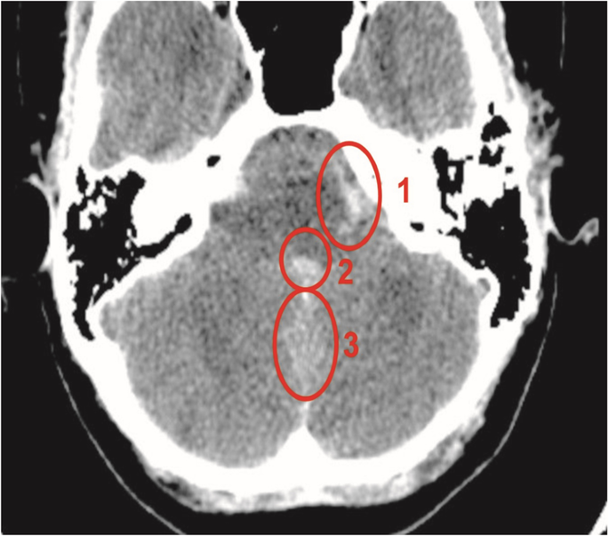
A 27-year-old female patient complained of sudden, severe headaches in the occipital region, nuchal rigidity, and drowsiness when she presented at our institution. A head computed tomography scan revealed intraventricular bleeding in the lateral and fourth ventricles with more extensive haemorrhaging in the frontal horns. A left posterior inferior cerebellar artery (PICA) aneurysm was confirmed via digital subtraction angiogram, and endovascular embolization was done. Two years later, the patient reported intense pain in the lower back along with symptoms suggestive of spinal cord compression. Spinal magnetic resonance imaging (MRI) showed spinal adhesions from C1 to L4, syringomyelia with some vasogenic oedema extending from T3 to T9 level, and a cyst in the lumbar region. Consequently, a right hemilaminectomy was performed along with microsurgical release of arachnoid adhesions and placement of a subdural drain. Radiological and symptomatic improvements were observed. Since then, the patient’s clinical condition has remained stable during the past three years of follow-up visits.
Conclusions
Literature on optimal treatment modalities and patient prognosis is scarce and debated. The time for symptom improvement depends on the level and extent of spinal cord involvement. Rehabilitation may be required for most patients, as complete symptomatic recovery may not be attainable.
中文翻译:
动脉瘤性蛛网膜下腔出血后迟发性脊髓蛛网膜炎伴脊髓空洞症:病例报告及患者经历
背景和重要性
与迟发性脊髓蛛网膜炎相关的脊髓空洞症或脊髓内充满液体的囊肿的形成是动脉瘤性蛛网膜下腔出血的罕见并发症。迄今为止,医学文献中已报道约 18 例病例,其中只有 2 例报道的患者年龄在 35 岁以下。
临床表现
一名 27 岁的女性患者在我们机构就诊时主诉枕部突然剧烈头痛、颈项强直和嗜睡。头部计算机断层扫描显示侧脑室和第四脑室有脑室内出血,额角有更广泛的出血。通过数字减影血管造影证实左小脑后下动脉(PICA)动脉瘤,并进行血管内栓塞。两年后,患者报告下背部剧烈疼痛,并伴有脊髓受压的症状。脊柱磁共振成像 (MRI) 显示从 C1 到 L4 的脊柱粘连、脊髓空洞症以及从 T3 延伸到 T9 水平的一些血管源性水肿,以及腰部区域的囊肿。因此,进行了右半椎板切除术,同时进行显微手术松解蛛网膜粘连并放置硬膜下引流管。观察到放射学和症状改善。此后,在过去三年的随访中,患者的临床状况一直保持稳定。
结论
关于最佳治疗方式和患者预后的文献很少且存在争议。症状改善的时间取决于脊髓受累的程度和程度。大多数患者可能需要康复,因为症状可能无法完全恢复。


 京公网安备 11010802027423号