Blood Cancer Journal ( IF 12.9 ) Pub Date : 2024-05-31 , DOI: 10.1038/s41408-024-01073-z Sheren Younes 1 , Ajay Subramanian 2 , Anum Khan 3 , Shuchun Zhao 1 , Michael Binkley 2 , Yasodha Natkunam 1
|
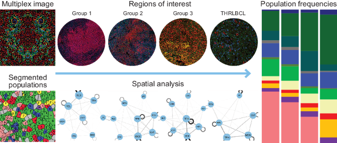
Nodular lymphocyte-predominant Hodgkin lymphoma (NLPHL) is a rare lymphoma with sparse tumor B-cells and a favorable prognosis. Variant growth patterns of NLPHL, however, often show advanced stage, progression to T-cell/histiocyte-rich large B-cell lymphoma (THRLBCL) and a worse prognosis. We studied the tumor microenvironment (TME) of NLPHL and THRLBCL using highplex imaging and spatial profiling at the single cell level. Our findings show distinct differences in TME composition and spatial configuration that differ among typical and variant NLPHL and THRLBCL. Typical NLPHL show abundant helper T-cell subsets, while THRLBCL show abundant cytotoxic T-cells and macrophages. Tumor B-cell size and content is lowest in typical NLPHL, followed by variant NLPHL, and highest in THRLBCL, whereas an opposite trend characterized TME B-cells. CD4/CD8 double-positive T-cells are seen in all NLPHL but not in the majority of THRLBCL and are spatially distant from LP-cells and TFH-rosettes. The differences in macrophage/monocyte content in distinguishing NLPHL pattern E from THRLBCL is further corroborated in independent cohorts of cases. Our results validate the current approach to classification and in addition provide novel insights that could be leveraged to refine clinical management for patients with this spectrum of lymphomas.
中文翻译:
结节性淋巴细胞为主的霍奇金淋巴瘤和富含 T 细胞/组织细胞的大 B 细胞淋巴瘤的空间表型
结节性淋巴细胞为主的霍奇金淋巴瘤 (NLPHL) 是一种罕见的淋巴瘤,肿瘤 B 细胞稀疏,预后良好。然而,NLPHL 的不同生长模式通常表现出晚期、进展为富含 T 细胞/组织细胞的大 B 细胞淋巴瘤 (THRLBCL) 和较差的预后。我们使用单细胞水平的高复合成像和空间分析研究了 NLPHL 和 THRLBCL 的肿瘤微环境 (TME)。我们的研究结果表明,典型和变异 NLPHL 和 THRLBCL 之间的 TME 组成和空间配置存在明显差异。典型的 NLPHL 显示丰富的辅助 T 细胞亚群,而 THRLBCL 显示丰富的细胞毒性 T 细胞和巨噬细胞。典型 NLPHL 中的肿瘤 B 细胞大小和含量最低,其次是变异型 NLPHL,THRLBCL 中最高,而 TME B 细胞则呈现相反的趋势。 CD4/CD8 双阳性 T 细胞在所有 NLPHL 中可见,但在大多数 THRLBCL 中不可见,并且在空间上远离 LP 细胞和 TFH 玫瑰花结。巨噬细胞/单核细胞含量在区分 NLPHL E 型和 THRLBCL 方面的差异在独立病例组中得到进一步证实。我们的结果验证了当前的分类方法,并提供了新的见解,可用于完善此类淋巴瘤患者的临床管理。


 京公网安备 11010802027423号