Scientific Reports ( IF 3.8 ) Pub Date : 2024-05-25 , DOI: 10.1038/s41598-024-62807-4
Linhui Yang 1, 2 , Kaige Wang 1, 2 , Weimin Li 1, 2 , Dan Liu 1, 2
|
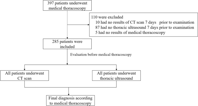
Septated pleural effusion is very common. The presence of septations in pleural effusion determines the local treatment strategy for such patients. Therefore, there is a pressing need for imaging techniques to assess the presence of septations. The objective of this research was to assess the diagnostic efficacy of computed tomography (CT) and chest ultrasound in identifying septated pleural effusion. We delineated the ultrasound and enhanced chest CT manifestations for diagnosing septated pleural effusions, and subsequently, we conducted a comparative analysis to assess the diagnostic efficacy of enhanced chest CT and ultrasound in identifying septated pleural effusions. Medical thoracoscopy served as the gold standard for confirming the diagnosis of septated pleural effusions. Ultrasound demonstrated a sensitivity of 82.6% (95% CI 73.3–89.7%) and a specificity of 100.0% (95% CI 98.1–NaN) for diagnosing septated pleural effusion. In comparison, enhanced chest CT exhibited a sensitivity of 59.8% (95% CI 49.0–69.9%) and a specificity of 87.0% (95% CI 81.5–91.4%). The positive predictive value for ultrasound was 100.0% (95% CI 95.3–100.0%), while for enhanced chest CT, it was 68.8% (95% CI 59.0–77.4%). Ultrasound yielded a negative predictive value of 92.3% (95% CI 87.5–NaN), and enhanced chest CT had a negative predictive value of 82.0% (95% CI 74.6–87.8%) in diagnosing septated pleural effusion. Thoracic ultrasound exhibits superior sensitivity and specificity compared to enhanced chest CT in diagnosing septated pleural effusions. Therefore, chest ultrasound is highly recommended as an adjunct for determining septated pleural effusion.
中文翻译:
胸部超声在识别胸膜疾病患者的分隔性积液方面优于CT
分隔性胸腔积液很常见。胸腔积液中是否存在分隔决定了此类患者的局部治疗策略。因此,迫切需要成像技术来评估隔膜的存在。本研究的目的是评估计算机断层扫描 (CT) 和胸部超声在识别分隔性胸腔积液方面的诊断效果。我们勾画了超声和增强胸部CT表现在诊断分隔性胸腔积液中的表现,随后进行了比较分析以评估增强胸部CT和超声在鉴别分隔性胸腔积液中的诊断效果。内科胸腔镜检查是确诊分隔性胸腔积液的金标准。超声诊断分隔性胸腔积液的敏感性为 82.6% (95% CI 73.3–89.7%),特异性为 100.0% (95% CI 98.1–NaN)。相比之下,增强胸部 CT 的敏感性为 59.8%(95% CI 49.0–69.9%),特异性为 87.0%(95% CI 81.5–91.4%)。超声检查的阳性预测值为 100.0% (95% CI 95.3–100.0%),而增强胸部 CT 的阳性预测值为 68.8% (95% CI 59.0–77.4%)。超声诊断分隔性胸腔积液的阴性预测值为 92.3% (95% CI 87.5–NaN),增强胸部 CT 的阴性预测值为 82.0% (95% CI 74.6–87.8%)。与增强胸部 CT 相比,胸部超声在诊断分隔性胸腔积液方面表现出更高的敏感性和特异性。因此,强烈建议胸部超声检查作为确定分隔性胸腔积液的辅助手段。


 京公网安备 11010802027423号