Cell Death and Differentiation ( IF 13.7 ) Pub Date : 2024-05-23 , DOI: 10.1038/s41418-024-01313-6 Konstantinos Kelepouras 1, 2 , Julia Saggau 1, 2, 3, 4 , Ana Beatriz Varanda 3, 4 , Matea Zrilic 2 , Christine Kiefer 1, 2 , Hassan Rakhsh-Khorshid 1, 2 , Ina Lisewski 2 , Iratxe Uranga-Murillo 5, 6 , Maykel Arias 5, 6 , Julian Pardo 5, 6 , Wulf Tonnus 7 , Andreas Linkermann 7, 8 , Alessandro Annibaldi 2 , Henning Walczak 3, 4, 9 , Gianmaria Liccardi 1, 2
|
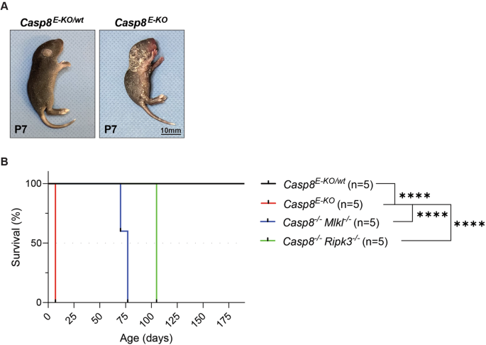
Necroptosis is a caspase-independent modality of cell death implicated in many inflammatory pathologies. The execution of this pathway requires the formation of a cytosolic platform that comprises RIPK1 and RIPK3 which, in turn, mediates the phosphorylation of the pseudokinase MLKL (S345 in mouse). The activation of this executioner is followed by its oligomerisation and accumulation at the plasma-membrane where it leads to cell death via plasma-membrane destabilisation and consequent permeabilisation. While the biochemical and cellular characterisation of these events have been amply investigated, the study of necroptosis involvement in vivo in animal models is currently limited to the use of Mlkl−/− or Ripk3−/− mice. Yet, even in many of the models in which the involvement of necroptosis in disease aetiology has been genetically demonstrated, the fundamental in vivo characterisation regarding the question as to which tissue(s) and specific cell type(s) therein is/are affected by the pathogenic necroptotic death are missing. Here, we describe and validate an immunohistochemistry and immunofluorescence-based method to reliably detect the phosphorylation of mouse MLKL at serine 345 (pMLKL-S345). We first validate the method using tissues derived from mice in which Caspase-8 (Casp8) or FADD are specifically deleted from keratinocytes, or intestinal epithelial cells, respectively. We next demonstrate the presence of necroptotic activation in the lungs of SARS-CoV-infected mice and in the skin and spleen of mice bearing a Sharpin inactivating mutation. Finally, we exclude necroptosis occurrence in the intestines of mice subjected to TNF-induced septic shock. Importantly, by directly comparing the staining of pMLKL-345 with that of cleaved Caspase-3 staining in some of these models, we identify spatio-temporal and functional differences between necroptosis and apoptosis supporting a role of RIPK3 in inflammation independently of MLKL versus the role of RIPK3 in activation of necroptosis.
中文翻译:
鼠磷酸化 MLKL-S345 原位检测对于体内坏死性凋亡评估的重要性
坏死性凋亡是一种不依赖半胱天冬酶的细胞死亡方式,与许多炎症病理有关。该途径的执行需要形成包含 RIPK1 和 RIPK3 的胞质平台,而 RIPK1 和 RIPK3 又介导假激酶 MLKL(小鼠中的 S345)的磷酸化。该刽子手的激活之后是其寡聚化并在质膜上积累,通过质膜不稳定和随后的透化导致细胞死亡。虽然这些事件的生化和细胞特征已得到充分研究,但动物模型体内坏死性凋亡的研究目前仅限于使用Mlkl -/-或Ripk3 -/-小鼠。然而,即使在许多模型中已经从遗传学角度证明了坏死性凋亡参与疾病病因学,但关于其中哪些组织和特定细胞类型受到影响的问题的基本体内特征仍然存在。致病性坏死性凋亡缺失。在这里,我们描述并验证了一种基于免疫组织化学和免疫荧光的方法,以可靠地检测小鼠 MLKL 在丝氨酸 345 (pMLKL-S345) 的磷酸化。我们首先使用小鼠组织验证该方法,其中 Caspase-8 (Casp8) 或 FADD 分别从角质形成细胞或肠上皮细胞中被特异性删除。接下来,我们证明了 SARS-CoV 感染小鼠的肺部以及带有 Sharpin 失活突变的小鼠的皮肤和脾脏中存在坏死性凋亡激活。最后,我们排除了 TNF 诱导的败血性休克小鼠肠道中坏死性凋亡的发生。 重要的是,通过直接比较其中一些模型中 pMLKL-345 的染色与裂解的 Caspase-3 染色的染色,我们确定了坏死性凋亡和细胞凋亡之间的时空和功能差异,支持 RIPK3 在独立于 MLKL 的炎症中的作用。 RIPK3 在坏死性凋亡激活中的作用。


 京公网安备 11010802027423号