Spinal Cord Series and Cases ( IF 0.7 ) Pub Date : 2024-05-23 , DOI: 10.1038/s41394-024-00650-5 Yushi Sakamoto , Takayoshi Shimizu , Bungo Otsuki , Shuichi Matsuda
|
Introduction
Spinal intradural arachnoid cysts (SIACs) are rare spinal entities that are categorized as primary or secondary pathologies. Secondary cysts can arise from various traumatic or inflammatory causes including subarachnoid hemorrhage, intrathecal injection or surgery, and infectious meningitis/arachnoiditis. Only a few cases of SIAC secondary to tuberculous meningitis have been previously reported, without details of the surgical treatment.
Case presentation
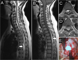
A 27-year-old woman diagnosed with tuberculous meningitis developed myelopathy caused by thoracic ventral SIAC and intradural abscess. The patient underwent abscess evacuation and cyst fenestration; however, cyst recurrence occurred. The 2nd surgery consisted of cyst resection via a posterolateral approach with expansive duraplasty and spinal arthrodesis. Re-recurrence occurred, and at the 3rd surgery, cyst-subarachnoid bypass was performed. One year after the 3rd surgery, the myelopathic symptoms recovered, and MR images demonstrated a decreased cyst size.
Discussion
Here, we report a rare case of recurrent thoracic SIAC secondary to tuberculous meningitis and arachnoiditis. Simple fenestration is associated with a high risk of recurrence in this pathology. Ventrally located thoracic cysts can be approached with posterolateral approach with pedicles resected followed by instrumented arthrodesis. Even in cases involving gross total resection of the cyst wall, there is a risk of recurrence. In such cases, cyst-subarachnoid bypass with a large-diameter tube can be effective.
中文翻译:
结核性脑膜炎继发复发性胸腹侧硬膜内蛛网膜囊肿的手术治疗一例报告
介绍
脊髓硬膜内蛛网膜囊肿 (SIAC) 是罕见的脊柱实体,可分为原发性或继发性病理。继发性囊肿可由各种创伤或炎症原因引起,包括蛛网膜下腔出血、鞘内注射或手术以及感染性脑膜炎/蛛网膜炎。此前仅报道了少数继发于结核性脑膜炎的SIAC病例,但未提供手术治疗的详细信息。
案例展示
一名 27 岁女性被诊断患有结核性脑膜炎,并因胸腹侧 SIAC 和硬膜内脓肿引起脊髓病。患者接受脓肿清除术和囊肿开窗术;然而,囊肿复发了。第二次手术包括通过后外侧入路囊肿切除术、扩张硬脑膜成形术和脊柱关节融合术。再次复发,第3次手术时进行了囊肿蛛网膜下腔搭桥术。第三次手术一年后,脊髓病症状恢复,磁共振图像显示囊肿尺寸缩小。
讨论
在此,我们报告一例继发于结核性脑膜炎和蛛网膜炎的复发性胸部 SIAC 的罕见病例。简单的开窗术与这种病理学复发的高风险相关。位于腹侧的胸椎囊肿可以采用后外侧入路,切除蒂,然后进行器械关节固定术。即使在囊肿壁完全切除的情况下,也存在复发的风险。在这种情况下,使用大直径管进行囊肿蛛网膜下腔搭桥术可能会有效。


 京公网安备 11010802027423号