Brain Structure & Function ( IF 2.7 ) Pub Date : 2024-05-04 , DOI: 10.1007/s00429-024-02799-z Wenhui Zhong 1, 2 , Qingwen Yang 1, 2 , Fenglan Wang 1, 2 , Xin Lin 1, 2 , Zhongqun Chen 1, 2 , Jing Xue 1, 2 , Wenna Zhao 1, 2 , Xiaoqing Liu 1, 2 , Bilin Rao 1, 2 , Jun Zhang 1, 2
|
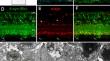
β-synuclein, a member of the synuclein family, is frequently co-expressed with α-synuclein in the neural system, where it serves to inhibit abnormal aggregation of α-synuclein in neurodegenerative diseases. Beyond its role in pathological conditions, β-synuclein plays various functions independently of α-synuclein. In our investigation, we discovered a broader expression of β-synuclein in the mouse retina compared to α-synuclein. This widespread pattern implies its potential significance in the retina. Through detailed examination via light- and electron-microscopic immunocytochemistry, we identified β-synuclein expression from the inner segment (IS) and outer segment (OS) of photoreceptor cells to the ganglion cell layer (GCL). Our findings unveiled unique features, including β-synuclein immunoreactive IS and OS of cones, higher expression in cone pedicles than in rod spherules, absence in horizontal cells, limited expression in cone bipolar dendrites and somas, higher expression in cone bipolar terminals, presence in most amacrine cells, and expression in almost majority of somas in GCL with an absence in intrinsically photosensitive retinal ganglion cell (ipRGCs) processes. Notably, all cholinergic amacrine cells express high β- but not α-synuclein, while dopaminergic amacrine cells express α-synuclein exclusively. These distinctive expression patterns offer valuable insights for further exploration into the functions of β-synuclein and its potential role in synuclein pathology within the retina.
中文翻译:
β-突触核蛋白在小鼠视网膜中的细胞特异性定位
β-突触核蛋白是突触核蛋白家族的一员,在神经系统中经常与 α-突触核蛋白共表达,可抑制神经退行性疾病中 α-突触核蛋白的异常聚集。除了在病理条件下的作用之外,β-突触核蛋白还独立于 α-突触核蛋白发挥各种功能。在我们的研究中,我们发现与 α-突触核蛋白相比,β-突触核蛋白在小鼠视网膜中的表达范围更广。这种广泛存在的模式意味着它在视网膜中的潜在重要性。通过光学和电子显微镜免疫细胞化学的详细检查,我们鉴定了从感光细胞的内段(IS)和外段(OS)到神经节细胞层(GCL)的β-突触核蛋白表达。我们的研究结果揭示了独特的特征,包括锥体的 β-突触核蛋白免疫反应性 IS 和 OS、锥体蒂中的表达高于杆球体、水平细胞中的缺失、锥体双极树突和体细胞中的有限表达、锥体双极末端中的较高表达、存在于大多数无长突细胞中表达,并且在 GCL 中的几乎大多数体细胞中表达,但内在光敏性视网膜神经节细胞 (ipRGC) 过程中不存在。值得注意的是,所有胆碱能无长突细胞均表达高水平的 β-突触核蛋白,但不表达 α-突触核蛋白,而多巴胺能无长突细胞仅表达 α-突触核蛋白。这些独特的表达模式为进一步探索 β-突触核蛋白的功能及其在视网膜内突触核蛋白病理学中的潜在作用提供了宝贵的见解。


 京公网安备 11010802027423号