Spinal Cord Series and Cases ( IF 0.7 ) Pub Date : 2024-04-25 , DOI: 10.1038/s41394-024-00644-3 Brian Fabian Saway , James Courtney , Jessica Barley , Bruce Frankel , Christoph Hofstetter , Stephen Kalhorn
|
Study design
Systematic review.
Objective
Contrast-enhanced ultrasound (CEUS) is an imaging modality that has only recently seen neurosurgical application. CEUS uses inert microbubbles to intraoperatively visualize vasculature and perfusion of the brain and spinal cord in real time. Observation and augmentation of spinal cord perfusion is vital component of the management of traumatic spinal cord injury, yet there are limited imaging modalities to evaluate spinal cord perfusion. CEUS provides an intraoperative imaging tool to evaluate spinal cord perfusion in real time. The objective of this review is to evaluate the current literature on the various applications and benefits of CEUS in traumatic spinal cord injury.
Setting
South Carolina, USA.
Methods
This review was written according to the PRISMA 2020 guidelines.
Results
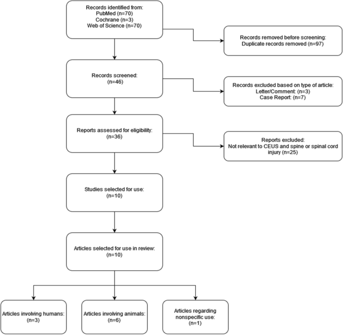
143 articles were found in our literature search, with 46 of them being unique. After excluding articles for relevance to CEUS and spinal cord injury, we were left with 10 papers. Studies in animal models have shown CEUS to be an effective non-invasive imaging modality that can detect perfusion changes of injured spinal cords in real time.
Conclusion
This imaging modality can provide object perfusion data of the nidus of injury, surrounding penumbra and healthy neural tissue in a traumatized spinal cord. Investigation in its use in humans is ongoing and remains promising to be an effective diagnostic and prognostic tool for those suffering from spinal cord injury.
中文翻译:
超声造影治疗创伤性脊髓损伤:当前和未来应用概述
学习规划
系统审查。
客观的
超声造影 (CEUS) 是一种最近才在神经外科领域得到应用的成像方式。 CEUS 使用惰性微泡在术中实时可视化大脑和脊髓的脉管系统和灌注。观察和增强脊髓灌注是治疗创伤性脊髓损伤的重要组成部分,但评估脊髓灌注的成像方式有限。 CEUS 提供了一种术中成像工具来实时评估脊髓灌注。本综述的目的是评估当前关于 CEUS 在创伤性脊髓损伤中的各种应用和益处的文献。
环境
美国南卡罗来纳州。
方法
本评论是根据 PRISMA 2020 指南撰写的。
结果
我们的文献检索共找到 143 篇文章,其中 46 篇是独特的。排除与 CEUS 和脊髓损伤相关的文章后,我们剩下 10 篇论文。动物模型研究表明,CEUS 是一种有效的非侵入性成像方式,可以实时检测受损脊髓的灌注变化。
结论
这种成像方式可以提供受伤脊髓中损伤病灶、周围半暗带和健康神经组织的对象灌注数据。其在人类中的使用研究正在进行中,并且仍然有希望成为脊髓损伤患者的有效诊断和预后工具。


 京公网安备 11010802027423号