Spinal Cord Series and Cases ( IF 0.7 ) Pub Date : 2024-04-17 , DOI: 10.1038/s41394-024-00639-0 Shuhei Ohtsubo , Masayuki Ohashi , Toru Hirano , Hideki Tashi , Tatsuo Makino , Keitaro Minato , Yusuke Mitsuma , Hiroyuki Deguchi , Rintaro Hoshino , Nobuko Ohashi , Kenta Furutani , Hiroyuki Kawashima , Kei Watanabe
|
Introduction
Although multimodal intraoperative neuromonitoring (IONM), which has high sensitivity and specificity, is typically performed during spinal deformity surgery, neurological status may deteriorate with delay after surgical maneuvers. Here, we report a rare case of delayed postoperative neurological deficit (DPND) that was not detected by IONM during posterior spinal fusion (PSF) for congenital scoliosis.
Case presentation
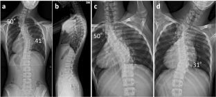
A 14-year-old male presented with congenital scoliosis associated with T3 and T10 hemivertebrae. Preoperative Cobb angle of proximal thoracic (PT) and main thoracic (MT) curves were 50° and 41°, respectively. PSF (T1-L1) without hemivertebrectomy was performed, and the curves were corrected to 31° and 21° in the PT and MT curves, respectively, without any abnormal findings in IONM, blood pressure, or hemoglobin level. However, postoperative neurological examination revealed complete loss of motor function. A revision surgery, release of the curve correction by removing the rods, was immediately performed and muscle strength completely recovered on the first postoperative day. Five days postoperatively, PSF was achieved with less curve correction (36° in the PT curve and 26° in the MT curve), without postoperative neurological deficits.
Discussion
Possible mechanisms of DPND in our patient are spinal cord ischemia due to spinal cord traction caused by scoliosis correction and spinal cord kinking by the pedicle at the concave side. Understanding the possible mechanisms of intra- and postoperative neural injury is essential for appropriate intervention in each situation. Additionally, IONM should be continued to at least skin closure to detect DPND observed in our patient.
中文翻译:
先天性脊柱侧凸后路脊柱融合术后迟发性截瘫:一例报告
介绍
尽管多模式术中神经监测(IONM)具有高灵敏度和特异性,通常在脊柱畸形手术期间进行,但神经状态可能会随着手术操作的延迟而恶化。在这里,我们报告了一个罕见的延迟性术后神经功能缺损 (DPND) 病例,在先天性脊柱侧凸的后路脊柱融合 (PSF) 过程中 IONM 未检测到该病例。
案例展示
一名 14 岁男性患有与 T3 和 T10 半椎骨相关的先天性脊柱侧凸。术前近胸廓(PT)和主胸廓(MT)曲线的Cobb角分别为50°和41°。进行 PSF(T1-L1),未进行半椎体切除,PT 和 MT 曲线分别校正为 31° 和 21°,IONM、血压或血红蛋白水平未发现任何异常。然而,术后神经系统检查显示运动功能完全丧失。立即进行了修复手术,即通过移除棒来解除曲线矫正,术后第一天肌肉力量完全恢复。术后五天,通过较少的曲线矫正(PT 曲线为 36°,MT 曲线为 26°)实现了 PSF,且术后无神经功能缺损。
讨论
本例患者发生 DPND 的可能机制是由于脊柱侧凸矫正引起的脊髓牵引和凹侧椎弓根的脊髓扭结导致脊髓缺血。了解术中和术后神经损伤的可能机制对于每种情况的适当干预至关重要。此外,IONM 应至少持续至皮肤闭合,以检测在我们的患者中观察到的 DPND。


 京公网安备 11010802027423号