Nature Catalysis ( IF 42.8 ) Pub Date : 2024-04-15 , DOI: 10.1038/s41929-024-01148-x
Teng-Xiang Huang , Xin Cong , Si-Si Wu , Jiang-Bin Wu , Yi-Fan Bao , Mao-Feng Cao , Liwen Wu , Miao-Ling Lin , Xiang Wang , Ping-Heng Tan , Bin Ren
|
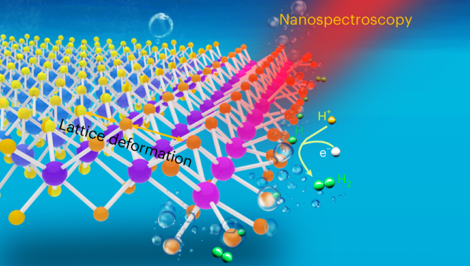
Understanding the structural evolution of individual active sites during a reaction is a long-standing target in surface science and catalysis. It is still challenging to precisely characterize in situ the intrinsic nature and evolution of the active site because the active site is too small for characterization techniques to decipher the local properties. Here we used electrochemical tip-enhanced Raman spectroscopy to monitor the geometric and electronic evolution of individual active sites of MoS2 during the hydrogen evolution reaction. Reconstruction regions of 40 nm with varied lattice and electron density from the edge to the nearby basal plane were observed during the hydrogen evolution reaction. We further revealed the progressive generation of active sites during the activation process. The synergistic reconstruction around edge due to the lattice deformation reduces the activation energy barriers and promotes the electrocatalytic reaction. These discoveries offer insights into our understanding of the active site and its dynamics during electrocatalysis.
中文翻译:
可视化电催化析氢反应过程中 MoS2 中各个活性位点的结构演化
了解反应过程中各个活性位点的结构演化是表面科学和催化领域的长期目标。由于活性位点太小,表征技术无法解读局部特性,因此精确地原位表征活性位点的内在性质和演化仍然具有挑战性。在这里,我们使用电化学尖端增强拉曼光谱来监测析氢反应过程中MoS 2各个活性位点的几何和电子演化。在析氢反应过程中,观察到从边缘到附近基面具有不同晶格和电子密度的 40 nm 重建区域。我们进一步揭示了激活过程中活性位点的逐渐生成。由于晶格变形而导致边缘周围的协同重构降低了活化能垒并促进了电催化反应。这些发现为我们了解电催化过程中的活性位点及其动力学提供了见解。


 京公网安备 11010802027423号