当前位置:
X-MOL 学术
›
Ann. N. Y. Acad. Sci.
›
论文详情
Our official English website, www.x-mol.net, welcomes your feedback! (Note: you will need to create a separate account there.)
Loose patch clamp membrane current measurements in cornus ammonis 1 neurons in murine hippocampal slices
Annals of the New York Academy of Sciences ( IF 4.1 ) Pub Date : 2024-04-11 , DOI: 10.1111/nyas.15123 Federico Bertagna 1, 2 , Shiraz Ahmad 2 , Rebecca Lewis 1, 2 , S Ravi P Silva 1, 3 , Johnjoe McFadden 1, 4 , Christopher L-H Huang 2, 5, 6 , Hugh R Matthews 5 , Kamalan Jeevaratnam 1, 2
Annals of the New York Academy of Sciences ( IF 4.1 ) Pub Date : 2024-04-11 , DOI: 10.1111/nyas.15123 Federico Bertagna 1, 2 , Shiraz Ahmad 2 , Rebecca Lewis 1, 2 , S Ravi P Silva 1, 3 , Johnjoe McFadden 1, 4 , Christopher L-H Huang 2, 5, 6 , Hugh R Matthews 5 , Kamalan Jeevaratnam 1, 2
Affiliation
|
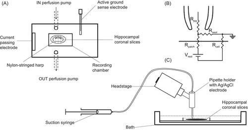
Hippocampal pyramidal neuronal activity has been previously studied using conventional patch clamp in isolated cells and brain slices. We here introduce the loose patch clamping study of voltage-activated currents from in situ pyramidal neurons in murine cornus ammonis 1 hippocampal coronal slices. Depolarizing pulses of 15-ms duration elicited early transient inward, followed by transient and prolonged outward currents in the readily identifiable junctional region between the stratum pyramidalis (SP) and oriens (SO) containing pyramidal cell somas and initial segments. These resembled pyramidal cell currents previously recorded using conventional patch clamp. Shortening the depolarizing pulses to >1–2 ms continued to evoke transient currents; hyperpolarizing pulses to varying voltages evoked decays whose time constants could be shortened to <1 ms, clarifying the speed of clamping in this experimental system. The inward and outward currents had distinct pharmacological characteristics and voltage-dependent inactivation and recovery from inactivation. Comparative recordings from the SP, known to contain pyramidal cell somas, demonstrated similar current properties. Recordings from the SO and stratum radiatum demonstrated smaller inward and outward current magnitudes and reduced transient outward currents, consistent with previous conventional patch clamp results from their different interneuron types. The loose patch clamp method is thus useful for in situ studies of neurons in hippocampal brain slices.
中文翻译:

小鼠海马切片中 Cornus ammonis 1 神经元的松散膜片钳膜电流测量
先前已使用传统膜片钳在分离细胞和脑切片中研究了海马锥体神经元活动。我们在这里介绍来自鼠角阿蒙尼斯 1 海马冠状切片中原位锥体神经元的电压激活电流的松散膜片钳研究。持续时间 15 毫秒的去极化脉冲引发了早期瞬态向内电流,随后在包含锥体细胞胞体和初始节段的锥体层 (SP) 和东方层 (SO) 之间易于识别的交界区域引发瞬态和长时间的向外电流。这些类似于先前使用传统膜片钳记录的锥体细胞电流。将去极化脉冲缩短至 >1–2 ms 会继续诱发瞬态电流;将超极化脉冲施加到不同的电压会引起衰变,其时间常数可以缩短至 <1 ms,从而阐明了该实验系统中的钳位速度。内向和外向电流具有不同的药理学特征以及电压依赖性失活和失活恢复。已知含有锥体细胞体的 SP 的比较记录显示出类似的电流特性。 SO 和辐射层的记录表明,向内和向外电流幅度较小,瞬态向外电流减少,这与之前不同中间神经元类型的传统膜片钳结果一致。因此,松散膜片钳方法可用于海马脑切片中神经元的原位研究。
更新日期:2024-04-11
中文翻译:
小鼠海马切片中 Cornus ammonis 1 神经元的松散膜片钳膜电流测量
先前已使用传统膜片钳在分离细胞和脑切片中研究了海马锥体神经元活动。我们在这里介绍来自鼠角阿蒙尼斯 1 海马冠状切片中原位锥体神经元的电压激活电流的松散膜片钳研究。持续时间 15 毫秒的去极化脉冲引发了早期瞬态向内电流,随后在包含锥体细胞胞体和初始节段的锥体层 (SP) 和东方层 (SO) 之间易于识别的交界区域引发瞬态和长时间的向外电流。这些类似于先前使用传统膜片钳记录的锥体细胞电流。将去极化脉冲缩短至 >1–2 ms 会继续诱发瞬态电流;将超极化脉冲施加到不同的电压会引起衰变,其时间常数可以缩短至 <1 ms,从而阐明了该实验系统中的钳位速度。内向和外向电流具有不同的药理学特征以及电压依赖性失活和失活恢复。已知含有锥体细胞体的 SP 的比较记录显示出类似的电流特性。 SO 和辐射层的记录表明,向内和向外电流幅度较小,瞬态向外电流减少,这与之前不同中间神经元类型的传统膜片钳结果一致。因此,松散膜片钳方法可用于海马脑切片中神经元的原位研究。


 京公网安备 11010802027423号