当前位置:
X-MOL 学术
›
J. Neurosci. Res.
›
论文详情
Our official English website, www.x-mol.net, welcomes your
feedback! (Note: you will need to create a separate account there.)
Alpha anteriorization and theta posteriorization during deep sleep
Journal of Neuroscience Research ( IF 2.9 ) Pub Date : 2024-04-02 , DOI: 10.1002/jnr.25325 Yue Cui 1 , Yu Li 1 , Qiqi Li 1 , Jing Huang 1, 2 , Xiaodan Tan 1, 2 , Chang'an A Zhan 1, 2, 3
Journal of Neuroscience Research ( IF 2.9 ) Pub Date : 2024-04-02 , DOI: 10.1002/jnr.25325 Yue Cui 1 , Yu Li 1 , Qiqi Li 1 , Jing Huang 1, 2 , Xiaodan Tan 1, 2 , Chang'an A Zhan 1, 2, 3
Affiliation

|
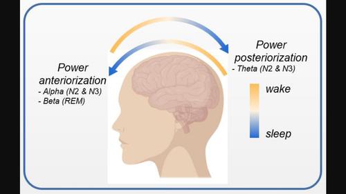
Brain states (wake, sleep, general anesthesia, etc.) are profoundly associated with the spatiotemporal dynamics of brain oscillations. Previous studies showed that the EEG alpha power shifted from the occipital cortex to the frontal cortex (alpha anteriorization) after being induced into a state of general anesthesia via propofol. The sleep research literature suggests that slow waves and sleep spindles are generated locally and propagated gradually to different brain regions. Since sleep and general anesthesia are conceptualized under the same framework of consciousness, the present study examines whether alpha anteriorization similarly occurs during sleep and how the EEG power in other frequency bands changes during different sleep stages. The results from the analysis of three polysomnography datasets of 234 participants show consistent alpha anteriorization during the sleep stages N 2 and N 3, beta anteriorization during stage REM , and theta posteriorization during stages N 2 and N 3. Although it is known that the neural circuits responsible for sleep are not exactly the same for general anesthesia, the findings of alpha anteriorization in this study suggest that, at macro level, the circuits for alpha oscillations are organized in the similar cortical areas. The spatial shifts of EEG power in different frequency bands during sleep may offer meaningful neurophysiological markers for the level of consciousness.
中文翻译:

深度睡眠期间的 α 前移和 θ 后移
大脑状态(觉醒、睡眠、全身麻醉等)与大脑振荡的时空动力学密切相关。先前的研究表明,脑电图 α 功率在通过异丙酚诱导进入全身麻醉状态后,从枕叶皮层转移到额叶皮层(α 前化)。睡眠研究文献表明,慢波和睡眠纺锤波是局部产生的,并逐渐传播到不同的大脑区域。由于睡眠和全身麻醉是在同一意识框架下概念化的,因此本研究检查了睡眠期间是否类似地发生了 α 前移,以及其他频段的脑电功率在不同睡眠阶段如何变化。对 234 名参与者的三个多导睡眠图数据集的分析结果显示,睡眠阶段 N2 和 N3 期间的 α 前移一致,REM 阶段的 β 前移,以及 N2 和 N3 阶段的 θ 后移。尽管已知负责睡眠的神经回路与全身麻醉并不完全相同,但本研究中 α 前移的结果表明,在宏观层面上,α 振荡的回路组织在相似的皮质区域中。睡眠期间不同频段的脑电功率空间偏移可能为意识水平提供有意义的神经生理学标志物。
更新日期:2024-04-02
中文翻译:

深度睡眠期间的 α 前移和 θ 后移
大脑状态(觉醒、睡眠、全身麻醉等)与大脑振荡的时空动力学密切相关。先前的研究表明,脑电图 α 功率在通过异丙酚诱导进入全身麻醉状态后,从枕叶皮层转移到额叶皮层(α 前化)。睡眠研究文献表明,慢波和睡眠纺锤波是局部产生的,并逐渐传播到不同的大脑区域。由于睡眠和全身麻醉是在同一意识框架下概念化的,因此本研究检查了睡眠期间是否类似地发生了 α 前移,以及其他频段的脑电功率在不同睡眠阶段如何变化。对 234 名参与者的三个多导睡眠图数据集的分析结果显示,睡眠阶段 N2 和 N3 期间的 α 前移一致,REM 阶段的 β 前移,以及 N2 和 N3 阶段的 θ 后移。尽管已知负责睡眠的神经回路与全身麻醉并不完全相同,但本研究中 α 前移的结果表明,在宏观层面上,α 振荡的回路组织在相似的皮质区域中。睡眠期间不同频段的脑电功率空间偏移可能为意识水平提供有意义的神经生理学标志物。


 京公网安备 11010802027423号