当前位置:
X-MOL 学术
›
Ceram. Int.
›
论文详情
Our official English website, www.x-mol.net, welcomes your
feedback! (Note: you will need to create a separate account there.)
Titanium dioxide bioceramics prepared by 3D printing method and its structure effect on stem cell behavior
Ceramics International ( IF 5.1 ) Pub Date : 2024-03-13 , DOI: 10.1016/j.ceramint.2024.03.164 Simeng Wang , Siqi Zhang , Yifan Cui , Xugang Lu , Mei Zhang , Jun Chen , Yipu Cao , Changchun Zhou , Bangcheng Yang
Ceramics International ( IF 5.1 ) Pub Date : 2024-03-13 , DOI: 10.1016/j.ceramint.2024.03.164 Simeng Wang , Siqi Zhang , Yifan Cui , Xugang Lu , Mei Zhang , Jun Chen , Yipu Cao , Changchun Zhou , Bangcheng Yang

|
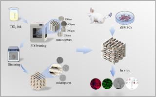
3D printing was widely used for the modification of bioceramic structures. Here, Bioactive titanium dioxide (TiO) ceramics with macropore structures were prepared by controlling the pore size through 3D printing, and further obtained secondary micropores by altering the sintering temperature. The structures and in vitro bioactivity of materials were characterized, and the properties of the ceramics for osteogenic differentiation of rabbits bone marrow mesenchymal stem cells (rBMSCs) were investigated. The results showed that titanium dioxide ceramics could induce apatite formation in SBF, which indicated it is bioactive. All materials exhibited porous structures, which could promote the proliferation and osteogenic differentiation of rBMSCs. Importantly, the structure with 200 μm pore size significantly promoted the proliferation and adhesion of cells. Furthermore, more secondary micropores formed on the surface of materials with the decreased sintering temperature, which promoted cell proliferation. The pore size of 400 μm significantly upregulated the expression of osteogenesis-related genes, including Col I, ALP, Runx2, and OPN. It had the strongest osteogenic ability when sintered at 1100 °C compared to other groups. This implies that the 3D-printed TiO ceramics have good osteogenic properties, and the hierarchical structure consisting of macroscopic pores and secondary micropores can control it well.
中文翻译:

3D打印方法制备二氧化钛生物陶瓷及其结构对干细胞行为的影响
3D打印被广泛用于生物陶瓷结构的修饰。在此,通过3D打印控制孔径来制备具有大孔结构的生物活性二氧化钛(TiO)陶瓷,并通过改变烧结温度进一步获得二次微孔。对材料的结构和体外生物活性进行了表征,并研究了陶瓷对兔骨髓间充质干细胞(rBMSCs)成骨分化的性能。结果表明,二氧化钛陶瓷可以在SBF中诱导磷灰石形成,这表明它具有生物活性。所有材料均表现出多孔结构,可以促进rBMSCs的增殖和成骨分化。重要的是,200μm孔径的结构显着促进细胞的增殖和粘附。此外,随着烧结温度的降低,材料表面形成更多的次生微孔,促进细胞增殖。 400μm孔径显着上调成骨相关基因的表达,包括Col I、ALP、Runx2和OPN。与其他组相比,在1100℃下烧结时其成骨能力最强。这意味着3D打印的TiO陶瓷具有良好的成骨性能,由宏观孔和次生微孔组成的分级结构可以很好地控制它。
更新日期:2024-03-13
中文翻译:

3D打印方法制备二氧化钛生物陶瓷及其结构对干细胞行为的影响
3D打印被广泛用于生物陶瓷结构的修饰。在此,通过3D打印控制孔径来制备具有大孔结构的生物活性二氧化钛(TiO)陶瓷,并通过改变烧结温度进一步获得二次微孔。对材料的结构和体外生物活性进行了表征,并研究了陶瓷对兔骨髓间充质干细胞(rBMSCs)成骨分化的性能。结果表明,二氧化钛陶瓷可以在SBF中诱导磷灰石形成,这表明它具有生物活性。所有材料均表现出多孔结构,可以促进rBMSCs的增殖和成骨分化。重要的是,200μm孔径的结构显着促进细胞的增殖和粘附。此外,随着烧结温度的降低,材料表面形成更多的次生微孔,促进细胞增殖。 400μm孔径显着上调成骨相关基因的表达,包括Col I、ALP、Runx2和OPN。与其他组相比,在1100℃下烧结时其成骨能力最强。这意味着3D打印的TiO陶瓷具有良好的成骨性能,由宏观孔和次生微孔组成的分级结构可以很好地控制它。


 京公网安备 11010802027423号