当前位置:
X-MOL 学术
›
Adv. Drug Deliver. Rev.
›
论文详情
Our official English website, www.x-mol.net, welcomes your feedback! (Note: you will need to create a separate account there.)
Advanced 3D imaging and organoid bioprinting for biomedical research and therapeutic applications
Advanced Drug Delivery Reviews ( IF 15.2 ) Pub Date : 2024-03-05 , DOI: 10.1016/j.addr.2024.115237 Sushila Maharjan , Chenshuo Ma , Bibhor Singh , Heemin Kang , Gorka Orive , Junjie Yao , Yu Shrike Zhang
Advanced Drug Delivery Reviews ( IF 15.2 ) Pub Date : 2024-03-05 , DOI: 10.1016/j.addr.2024.115237 Sushila Maharjan , Chenshuo Ma , Bibhor Singh , Heemin Kang , Gorka Orive , Junjie Yao , Yu Shrike Zhang
|
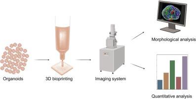
Organoid cultures offer a valuable platform for studying organ-level biology, allowing for a closer mimicry of human physiology compared to traditional two-dimensional cell culture systems or non-primate animal models. While many organoid cultures use cell aggregates or decellularized extracellular matrices as scaffolds, they often lack precise biochemical and biophysical microenvironments. In contrast, three-dimensional (3D) bioprinting allows precise placement of organoids or spheroids, providing enhanced spatial control and facilitating the direct fusion for the formation of large-scale functional tissues . In addition, 3D bioprinting enables fine tuning of biochemical and biophysical cues to support organoid development and maturation. With advances in the organoid technology and its potential applications across diverse research fields such as cell biology, developmental biology, disease pathology, precision medicine, drug toxicology, and tissue engineering, organoid imaging has become a crucial aspect of physiological and pathological studies. This review highlights the recent advancements in imaging technologies that have significantly contributed to organoid research. Additionally, we discuss various bioprinting techniques, emphasizing their applications in organoid bioprinting. Integrating 3D imaging tools into a bioprinting platform allows real-time visualization while facilitating quality control, optimization, and comprehensive bioprinting assessment. Similarly, combining imaging technologies with organoid bioprinting can provide valuable insights into tissue formation, maturation, functions, and therapeutic responses. This approach not only improves the reproducibility of physiologically relevant tissues but also enhances understanding of complex biological processes. Thus, careful selection of bioprinting modalities, coupled with appropriate imaging techniques, holds the potential to create a versatile platform capable of addressing existing challenges and harnessing opportunities in these rapidly evolving fields.
中文翻译:

用于生物医学研究和治疗应用的先进 3D 成像和类器官生物打印
类器官培养为研究器官水平生物学提供了一个有价值的平台,与传统的二维细胞培养系统或非灵长类动物模型相比,可以更接近地模拟人类生理学。虽然许多类器官培养物使用细胞聚集体或脱细胞细胞外基质作为支架,但它们通常缺乏精确的生化和生物物理微环境。相比之下,三维(3D)生物打印可以精确放置类器官或球体,提供增强的空间控制并促进直接融合以形成大规模功能组织。此外,3D生物打印能够微调生化和生物物理线索,以支持类器官的发育和成熟。随着类器官技术的进步及其在细胞生物学、发育生物学、疾病病理学、精准医学、药物毒理学和组织工程等不同研究领域的潜在应用,类器官成像已成为生理和病理研究的重要方面。这篇综述强调了成像技术的最新进展,这些技术对类器官研究做出了重大贡献。此外,我们讨论了各种生物打印技术,强调它们在类器官生物打印中的应用。将 3D 成像工具集成到生物打印平台中可以实现实时可视化,同时促进质量控制、优化和全面的生物打印评估。同样,将成像技术与类器官生物打印相结合可以为组织形成、成熟、功能和治疗反应提供有价值的见解。 这种方法不仅提高了生理相关组织的再现性,而且增强了对复杂生物过程的理解。因此,仔细选择生物打印方式,再加上适当的成像技术,有可能创建一个多功能平台,能够应对这些快速发展的领域中现有的挑战并利用机遇。
更新日期:2024-03-05
中文翻译:
用于生物医学研究和治疗应用的先进 3D 成像和类器官生物打印
类器官培养为研究器官水平生物学提供了一个有价值的平台,与传统的二维细胞培养系统或非灵长类动物模型相比,可以更接近地模拟人类生理学。虽然许多类器官培养物使用细胞聚集体或脱细胞细胞外基质作为支架,但它们通常缺乏精确的生化和生物物理微环境。相比之下,三维(3D)生物打印可以精确放置类器官或球体,提供增强的空间控制并促进直接融合以形成大规模功能组织。此外,3D生物打印能够微调生化和生物物理线索,以支持类器官的发育和成熟。随着类器官技术的进步及其在细胞生物学、发育生物学、疾病病理学、精准医学、药物毒理学和组织工程等不同研究领域的潜在应用,类器官成像已成为生理和病理研究的重要方面。这篇综述强调了成像技术的最新进展,这些技术对类器官研究做出了重大贡献。此外,我们讨论了各种生物打印技术,强调它们在类器官生物打印中的应用。将 3D 成像工具集成到生物打印平台中可以实现实时可视化,同时促进质量控制、优化和全面的生物打印评估。同样,将成像技术与类器官生物打印相结合可以为组织形成、成熟、功能和治疗反应提供有价值的见解。 这种方法不仅提高了生理相关组织的再现性,而且增强了对复杂生物过程的理解。因此,仔细选择生物打印方式,再加上适当的成像技术,有可能创建一个多功能平台,能够应对这些快速发展的领域中现有的挑战并利用机遇。


 京公网安备 11010802027423号